Novel nucleoside analogs and use thereof in therapeutic treatment

a technology of nucleoside analogs and nucleosides, applied in the field of new nucleoside analogs and their use in therapeutic treatment, can solve the problems of rapid progression to death, lack of complete efficacy of mouse models, and inability to fully achieve the effect of mouse models

Pending Publication Date: 2019-07-25
CORNELL UNIVERSITY
View PDF1 Cites 0 Cited by
  • Summary
  • Abstract
  • Description
  • Claims
  • Application Information

AI Technical Summary

Benefits of technology

[0180]BC3 and U266 cells were seeded at a density of 0.5 million cells / ml and treated with DMSO only, 1 μM or 1.0 μM 6-ETI for BC3 cells and 0.5 μM 6-ETI for U266 cells for 24 hrs. At 24 hrs post 6-ETI treatment, 0.5 million cells of each of the treated conditions were labeled with 10 μM of EdU for 2 hours then fixed for 30 min using 2% paraformaldehyde supplemented with 0.3M sucrose. Cells were then washed in PBS and perineabilized with 0.2% saponin, PBS with 1% BSA and 0.3M sucrose for 20 min at room temperature. During this incubation time, a click-iT cocktail mix containing, click iT reaction buffer, CuSO4, alexafluor488 and click iT reaction buffer additive was prepared according to the manufacturer's instructions. Cells were washed in PBS after permeabilization and the click-iT cocktail mix was added for 30 min at room temperature in the absence of light. To visualize DNA, cells were counterstained with Hoechst (1:1000) for another 30 min then washed and resuspended in PBS with 1% BSA. 20 μl of the resuspended cells were combined with 20 μl of mounting media Fluoromount-G (Southern biotech) and cells were mounted on Ibidi μ-slides. Cells were imaged using DeltaVision image restoration microscope (Rockefeller center).
[0181]1*105 treated cells were pelleted into a 96-well round bottom plate. Cells were washed once with PBS, and fixed in PBS+2% paraformaldehyde+0.03M sucrose for 15 min at room temperature. Cells were then washed twice with PBS+1% BSA+0.03M sucrose, permeabilized with PBS+0.2% saponin+1% BSA+0.03M sucrose (IFA Wash) for 15 min at room temperature, and blocked for 1 hour in PBS+5% normal goat serum. Cells were then stained in IFA Wash containing phospho-histone-H2A.X (ser139) antibody (γ-H2AX) at 1:100 dilution overnight at 4° C. Following two washes with PBS+1% BSA+0.03M sucrose, cells were re-permeabilized and re-blocked as above prior to incubation for 1 hour at room temperature with anti-rabbit-AlexaFluor594 Hoechst diluted 1:1000 in IFA Wash. Cells were then washed twice with PBS+1% BSA+0.03M sucrose, resuspended in 30 μl Fluoromount-G (Southern Biotech) and transferred to 16-well fbidi μ-slides for imaging. Images were taken on an Applied Precision Deltavision Image Restoration Microscope using Softworx analysis software. Z-stacks with a 0.2 μm step size were taken at 100× magnification. Stacks were subjected to deconvolution analysis and projections were made superimposing 5 representative z-planes to generate the final image.
[0182]A mixture of ethanethiol (26 g, 419 mmol), methanol (16 ml), and sodium hydroxide (6.7 ml, 20% aqueous solution) was cooled to 0° C., using an ice water bath. The mixture was stirred for 30 minutes, at which point 6-chloropurine riboside (2 g, 7 mmol) was added. The resulting solution was slowly warmed to ambient temperature and stirred for 12 hours, then was neutralized with glacial acetic acid (2.4 ml). Solvent was then removed using rotary evaporator; a small amount of bleach was placed in the solvent trap to quench any remaining ethanethiol. The resulting crude product was dissolved in acetone (100 mL) and then passed through a short Celite plug to remove minor impurities. Concentration in vacuo provided 6-ETI as a white powder (2.11 g, 6.7 mmol, 96%). 1H NMR (500 MHz, CD3OD) δ 8.69 (s, 1H), 8.59 (s, 1H), 6.09 (d, J=5.8 Hz, 1H), 4.75 (dd, J=5.5, 5.4 Hz, 1H), 4.37 (dd, J=5.1, 3.3 Hz, 1H), 4.18 (dd, J=6.2, 3.1 Hz, 1H), 3.91 (dd, J=12.4, 2.9 Hz, 1H), 3.78 (dd, J=12.4, 3.1 Hz, 1H), 3.42 (q, J=7.4 Hz, 2H), 1.45 (t, J=7.4 Hz, 3H).
[0183]Exponentially growing BC3-derived reporter cell lines (BC3NFκB-luc#6 or BC3NFRen-luc#3), and unrelated luciferase expressing U251-pGL3 cells were resuspended in respective RPMI-1640 complete media, and plated in a 96-well tissue culture microplate at 7.5*105 cells / mL and 3*105 cells / mL, respectively, in the presence or absence of varying concentrations of test compounds. After 24 hours of incubation, the luciferase activity was measured using Steady-Glo Luciferase Assay System or Dual-Glo Luciferase assay system (Promega, Madison, Wis.), according to the manufacturer's instructions.
[0184]BC3 cells in the exponential phase were plated at 2*105 cells / mL, and treated with 100 nM, 1 μM, or 10 μM 6-ETI for 6, 24, or 48 hours. Expression of viral latent and lytic genes was determined using qRT-PCR as previously described (Soulier J. et al., Blood. 1995;86(4):1276-80).
[0185]Lytic reactivation low was performed as previously described (Soulier J. et al., Blood 1995;86(4):1276-80). JSC1 B3.1 cells in the exponential phase were seeded at 2*105 cells / mL and treated with 1 or 10 μM 6-ETI for 24 or 48 hours, or 20 ng / mL lytic inducer TPA for 48 hours.

Problems solved by technology

However, PEL itself remains by large a highly aggressive and intractable disease, with rapid progression to death.
Therefore, the need for specific and effective therapeutics for diseases involving KSHV is pressing, albeit challenging due to the latent nature of the virus.
However, none of these have been completely effective in mouse models or involved the use of compounds that are already past early phases of clinical trials.

Method used


Image

  • Novel nucleoside analogs and use thereof in therapeutic treatment
  • Novel nucleoside analogs and use thereof in therapeutic treatment
  • Novel nucleoside analogs and use thereof in therapeutic treatment

Examples


example 1

and Methods

Electrophoretic Mobility Shift Assays

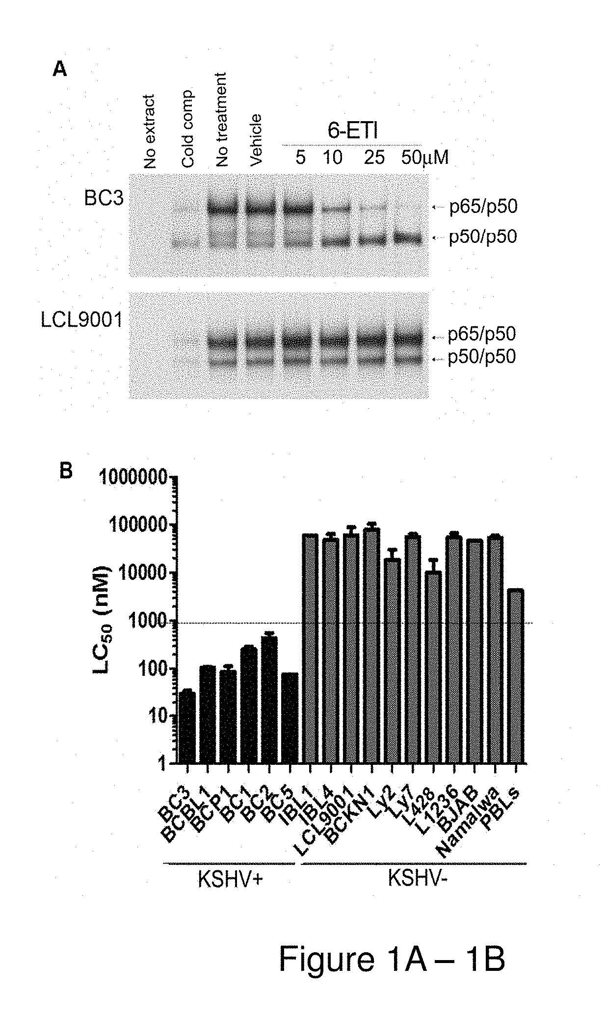
[0159]BC3 or LCL9001 cells were plated at 5*105 cells / ml, and treated with the hit compounds at 25 μM or with NSC39368 at 5, 10, 25 and 50 μM for 24 hours. Nuclear extracts were obtained as previously described (Keller S. A. et al., Blood. 2000 96(7):2537-42), and electrophoretic mobility shift assays (EMSAs) were performed using an NF-κB-specific oligonucleotide probe.

Viability Assays

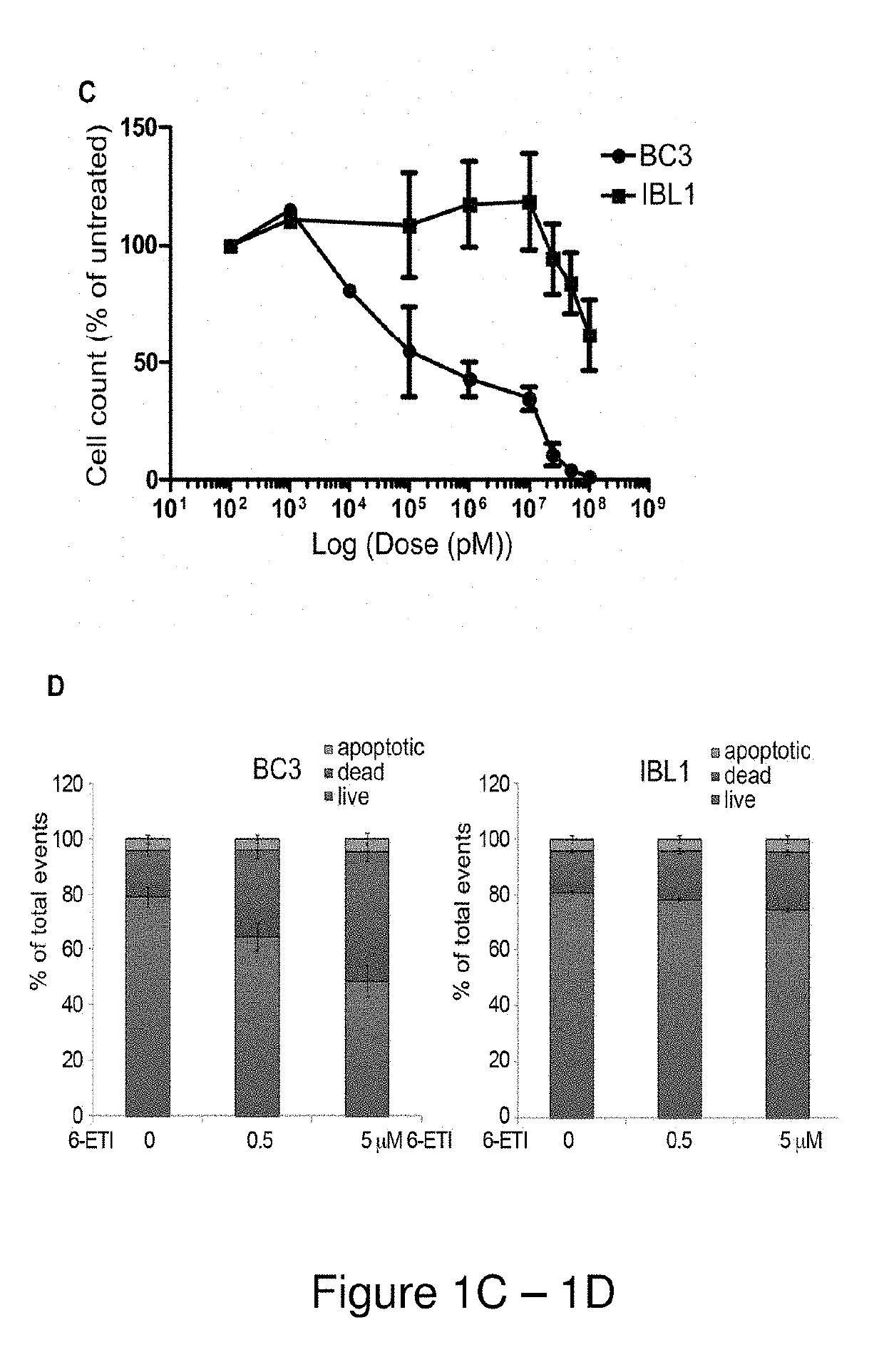
[0160]Cell viability assays were performed by plating log-phase cells in RPMI complete media with 20% FBS at 1*105 cells / mL, with concentrations of 6-ETI or phospho-6-ETI ranging from 0.1 nM to 100 μM. ATP content at 24, 48 and 72 hours post-treatment was measured by the CellTiter-Glo kit (Promega, Madison, Wis.), or by Trypan blue assay as indicated. The LC50 for 6-ETI in each cell line was determined using online EC50 software at changbioscience.com or using GraphPad Prism to determine the LC50 for 6-ETI, phospho-6-ETI and nucleoside analogs in MM cell ...

example 2

nt of a High Throughput-Screening Assay Using a PEL-Specific NF-κB Reporter Cell Line

[0189]The PEL cell line BC3 was previously modified through transduction of NFκB-firefly luciferase plasmid, followed by selection and subcloning, to yield the BC3NFκB-luc#6 single reporter cell line (Keller S A et al, Blood, 2006;107(8):3295-302), The Training Set (230 compounds) was screened using the BC3NFκB-luc#6 single reporter cell line, and optimal assay conditions for high-throughput screening (HTS) were determined. The established cell-based luciferase reporter assay was used to screen the NIH Diversity Set (1981 compounds) at 5 μM, yielding 60 primary hits that demonstrated at least 50% NFκB-luciferase inhibition, for an initial hit rate of 3%. Re-testing of these hit compounds determined 50 to be genuine. p As a next step to determine specificity, the inventors developed a secondary assay, wherein the single reporter cell line was lentivirally transduced with a constitutive renilla lucife...

example 3

of 6-ETI

[0190]The inventors examined the six hit compounds obtained at the end of HTS for inhibition of the NF-κB pathway in parental BC3 cells. The compound NSC39368 effectively inhibited active heterodimer p65 / p50 binding to the NF-κB response element by electrophoretic mobility shift assay (EMSA). This inhibition occurred in the BC3 PEL cell line, but not in a control lymphoblastoid cell line (LCL9001) containing EBV. This result identified NSC39368 or 6-ethylthioinosine, a nucleoside analog, hereafter referred to as 6-ETI, as the most promising compound for further investigation.

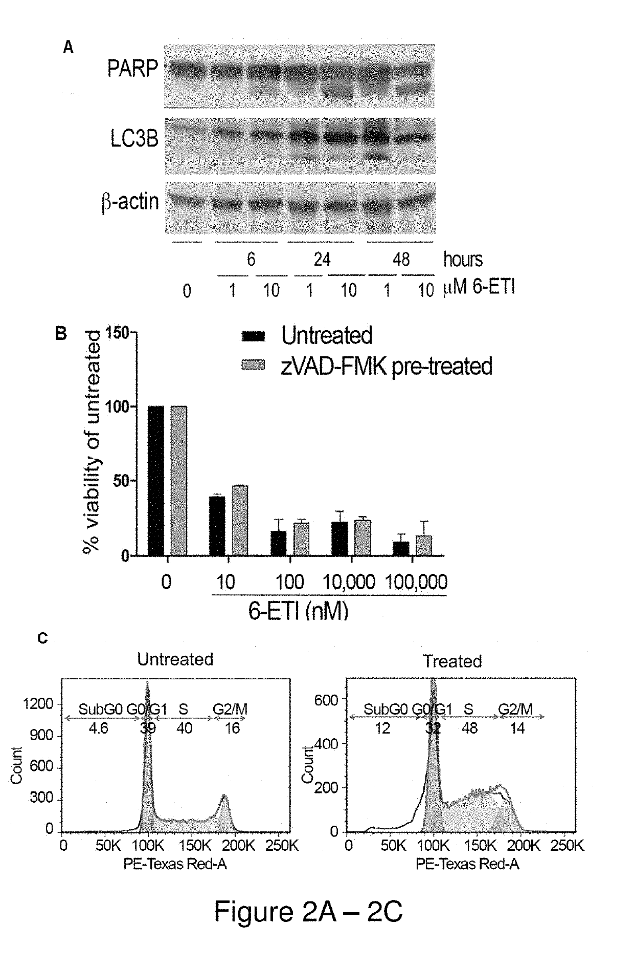
[0191]NF-κB reporter assays performed on BC3NFRen-luc#3 cells confirmed that 6-ETI inhibited NF-κB activity in this cell line as early as 6 hours post-treatment. 6-ETI treatment in BC3 cells was found to decrease levels of active subunit p65 in the nucleus, while leaving inactive subunit p50 unchanged, and it also decreased levels of IL6, an NF-κB dependent gene in PEL cells. Other components of classica...


PUM

PropertyMeasurementUnit
wavelengthaaaaaaaaaa
voltageaaaaaaaaaa
temperatureaaaaaaaaaa

Abstract

The present disclosure is directed to novel nucleoside analog compounds and methods for treating diseases characterized by high expression levels of adenosine kinase (ADK).

Description

CROSS REFERENCE TO RELATED APPLICATION[0001]This application claims the benefit of priority from U.S. Provisional Application No. 62 / 323,637, filed Apr. 16, 2016, the entire contents of which are incorporated herein by reference.STATEMENT REGARDING FEDERALLY SPONSORED RESEARCH OR DEVELOPMENT[0002]This invention was made with government support under Grant Nos. NIH / NCI 1R01CA15422, NIH / NCI N01-CO-12400 and NIH T32-AI007621, awarded by the National Institutes of Health and the National Cancer Institute. The government has certain rights in the invention.BACKGROUND[0003]The γ-herpesvirus KSHV, also called HHV-8, is the etiologic agent of Kaposi's sarcoma (KS) (Chang Y. et al., Science. 1994; 266(5192):1865-9), multicentric Castleman's disease (MCD) (Soulier J. et al., Blood. 1995;86(4):1276-80.), and primary effusion lymphoma (PEL) (Cesarman E. et al., N Engl J Med. 1995;332(18):1186-91). KS, the most common malignancy in AIDS patients, is a tumor of endothelial origin, while PEL is a ...

Claims


Application Information

Patent Timeline
Patent Type & Authority Applications(United States)
IPC IPC(8): C07H19/16C12Q1/6886G01N33/574G01N33/50A61P35/00
CPCC07H19/16C12Q1/6886G01N33/574G01N33/5011A61P35/00C12Q2600/106C12Q2600/158G01N2333/912C07H19/167C07H19/20C07H19/213
Inventor CESARMAN, ETHELNAYAR, UTTHARAWARREN, J. DAVIDSADEK, JOULIANA
Owner CORNELL UNIVERSITY
Why Eureka
Try Eureka