STEM CELL MICROPARTICLES AND miRNA

a technology of stem cells and microparticles, which is applied in the field of stem cell microparticles and mirna, can solve the problems of low regenerative capacity of damaged central nervous system (cns) tissue, no convincing evidence for direct long-term effect of transplanted stem cells, and potential safety risks of tumour or ectopic tissue formation, so as to reduce migration, promote differentiation of tumours, and reduce the effect of stem cell marker nestin

Pending Publication Date: 2019-12-19
RENEURON LTD
View PDF0 Cites 2 Cited by
  • Summary
  • Abstract
  • Description
  • Claims
  • Application Information

AI Technical Summary

Benefits of technology

[0019]The Examples include a pilot in vivo study of the administration of microparticles of the invention to human glioblastoma xenografts, observing tumour sensitivity to the microparticles, a trend towards a reduction in tumour volume, and increased survival. Histopathology of the tumour cells shows, in one animal, a particularly dramatic and effective ablation of the tumour mass.
[0031]The therapy may also be a prophylactic therapy to induce tolerance, typically immunotolerance, in a host that is subsequently, concurrently or simultaneously to receive the stem cells from which the microparticle is derived. The administration of one or more doses of microparticles of the invention to a patient, prior to or concurrent with administration of a stem cell therapy, can be used to reduce the risk of an adverse immune response, i.e. “rejection”, of the stem cell therapy.
[0033]It has also been found that it is possible to alter the production of microparticles by stem cells, by culturing the stem cells (optionally for at least 10 weeks) and adding components to the culture medium, by culturing the stem cells (optionally for at least 10 weeks) under hypoxic conditions, or by co-culture with other cell types (optionally for at least ten weeks), thereby providing an improved method of producing stem cell microparticles.

Problems solved by technology

However, there are drawbacks to the use of stem cells in therapy: there is a need for a consistent and substantial supply of stem cells with functional and phenotypic stability and the associated high costs and time delay caused by cell generation, storage, transport and handling; there is a requirement for immunological compatibility to avoid rejection of the stem cells by the recipient; and there are complex regulatory issues related to potential safety risks of tumour or ectopic tissue formation.
Further, despite the therapeutic efficacy of stem cell transplantation, there is no convincing evidence for a direct long-term effect of the transplanted stem cells, for example through engraftment and differentiation into reparative or replacement cells.
Damaged central nervous system (CNS) tissue has very limited regenerative capacity so that loss of neurological function is often chronic and progressive.
Production of human MSCs is limited by the inability of these cells to expand in numbers stably beyond approximately 15-20 population doublings.
Whereas some of the drawbacks of using stem cells directly as therapeutic agents are overcome by using the mesenchymal stem cell-derived exosomes (e.g. storage, transport and handling), the problem remains of providing a consistent and substantial supply of functionally and phenotypically stable stem cells to produce the exosomes.
In the absence of a stem cell line, replenishment of the cells through repeated derivation from a source of stem cells is required, which incurs recurring costs for testing and validation of each new batch.
Furthermore, the diseases and disorders that can be treated by MSCs may be limited.

Method used


Image

  • STEM CELL MICROPARTICLES AND miRNA
  • STEM CELL MICROPARTICLES AND miRNA
  • STEM CELL MICROPARTICLES AND miRNA

Examples


example 1

mes that Inhibit Cell Migration

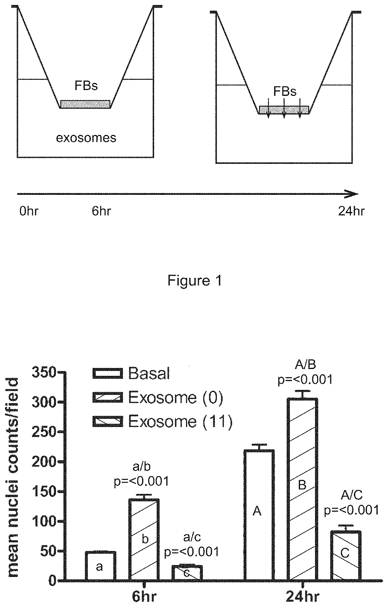
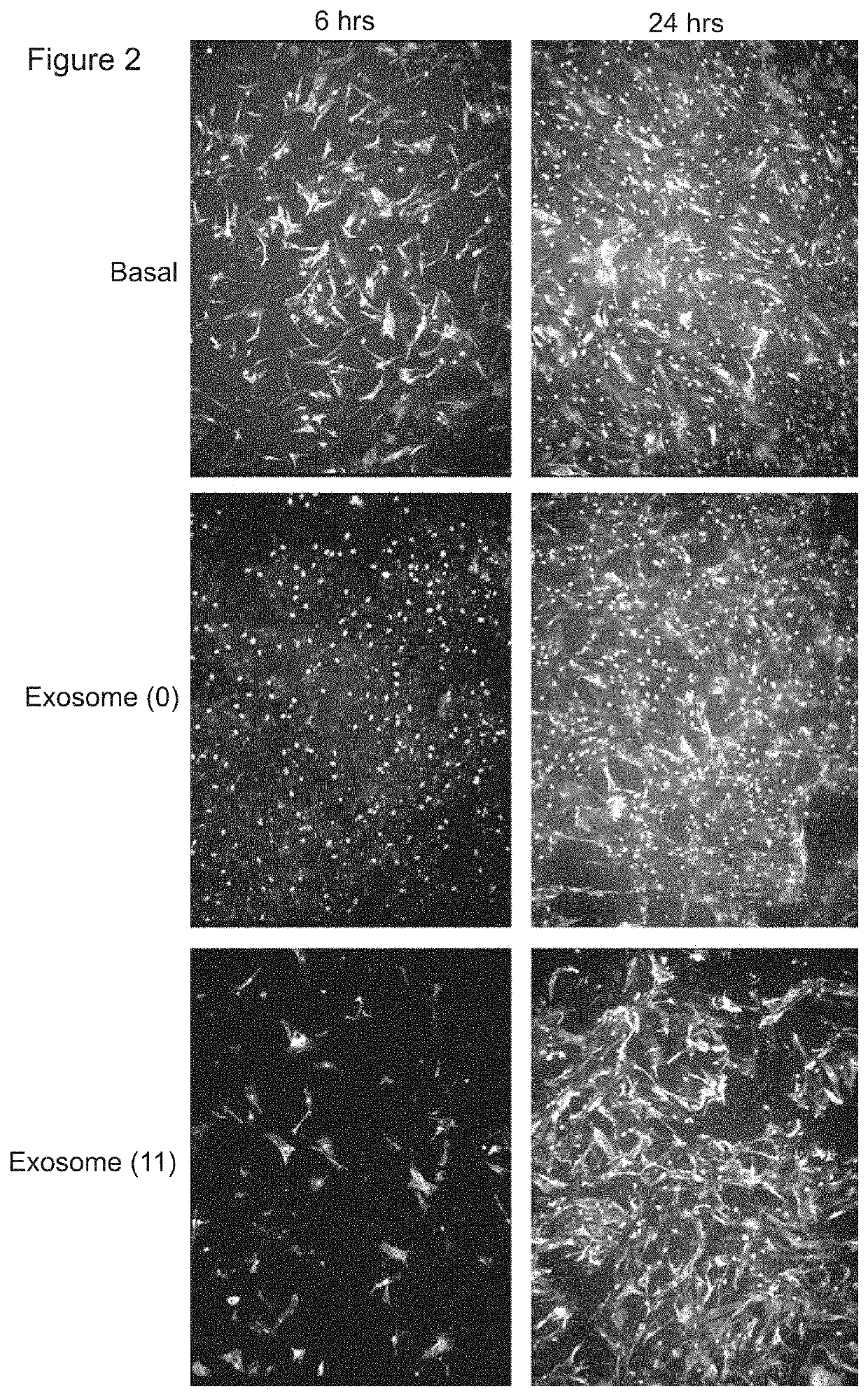
[0366]A transwell assay was used to study the migratory response of human dermal fibroblasts to different populations of exosomes. Experiments were performed in triplicate. 200,000 human dermal fibroblast cells (“FBs”) were placed on the upper layer of a cell permeable membrane (8 μm pore size; 24-well plate) and a solution (basal medium) containing or lacking 20 μg / ml exosomes was placed in contact with the underside of the cell permeable membrane (FIG. 1, top panel). The exosomes were collected from CTX0E03 cells cultured for 0 weeks (“0”) or 11 weeks (“11”) in an Integra CeLLine AD1000 multi-chamber bioreactor. Following an incubation period (6 or 24 hours; control: 0 hours), the human dermal fibroblast cells that migrated through the membrane were stained (using a fluorescent-dye conjugated anti-actin antibody and Hoechst Fluorescent Stain for nuclei) and counted (six random microscope fields per sample) as an indicator of the cells' migratory resp...

example 2

Isolated from the Medium of NSCs Cultured for 2 or 6 Weeks Promote Fibroblast Migration

Method—

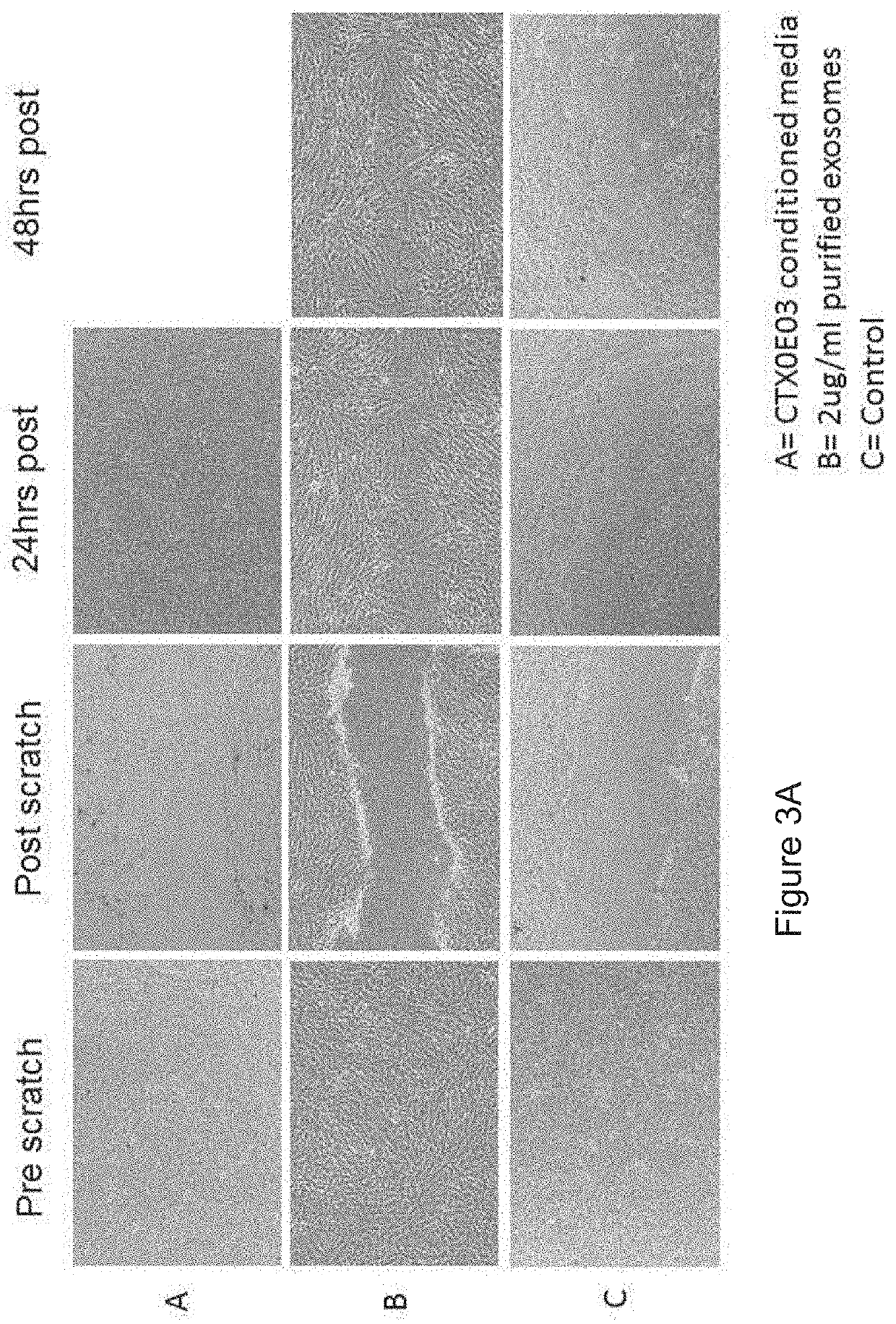
[0371]Wound closure / scratch assay[0372]Seed 0.25×10⁶ NHDF (normal human dermal fibroblasts) per well of a 12 well plate and allow to become confluent (24 hours)[0373]Remove growth factors for 24 hrs[0374]Remove cells (scratch) and incubate with exosomes / conditioned media[0375]Image effected area over 48 hrs[0376]Estimate area using Image J

Results

[0377]

TABLE 2Wound closure / scratch assay representing the migration activity of normal human dermal fibroblasts (NHDF) cultured in CTX0E03 conditioned media or upon the addition of purified exosomes.Wound closure (%)0 h24 h48 hCTX0E03 conditioned media 0% 100%2 ug / ml exosomes0%95.4% 100%Control0%48.1%49.7%

[0378]Wound closure was calculated as the area covered by cells in relation to the initial wound area, as determined at 0 h. Wound closure is expressed as the percentage of the initial wound area at time 0 h. These data are also shown, photographic...

example 3

oma Engraftment Assay—Destruction of Tumour Cells

[0384]U373 glioblastoma cells were pre-treated in vitro for 24 hours with exosomes isolated from CTX0E03 cells cultured for 11 weeks in an Integra CeLLine bioreactor before implantation into the striatum of Balb-C mice brains.

[0385]As shown in FIG. 19, the exosome-treated glioblastoma cells did not engraft into the striatum. Histopathology demonstrated the presence of necrotic U373 cell bodies at the site of implantation and evidence of gliosis—a host cellular immune response.

[0386]These data suggest utility of these exosomes in the treatment of cancer, by promoting the destruction of a tumour by the immune system, particularly a tumour of the CNS such as a glioblastoma.


PUM

PropertyMeasurementUnit
sizeaaaaaaaaaa
sizeaaaaaaaaaa
sizeaaaaaaaaaa

Abstract

This invention relates to stem cell microparticles and miRNA isolated from these microparticles, their use and production thereof, in particular neural stem cell microparticles and their use in therapy of cancer, typically a nestin-positive cancer. The cancer may be glioma, melanoma, breast cancer, pancreatic cancer or prostate cancer. The stem cell microparticle is typically an exosome or microvesicle and may be derived from a neural stem cell line. The neural stem cell line may be a conditionally-immortalised stem cell line such as CTX0E03 (deposited at the ECACC with Accession No. 04091601).

Description

CROSS-REFERENCES TO RELATED APPLICATIONS[0001]This application is continuation of U.S. patent application Ser. No. 15 / 027,424, filed Apr. 5, 2016, which is a national stage application under 35 U.S.C. § 371 of International Application No. PCT / GB2014 / 053044, filed Oct. 9, 2014, which in turn claims priority to United Kingdom Patent Application No. 1317887.6, filed Oct. 9, 2013, and International Application No. PCT / GB2014 / 052509, filed Aug. 14, 2014, the content of each of which is hereby incorporated by reference into this application in its entirety.REFERENCE TO A “SEQUENCE LISTING,” SUBMITTED AS ASCII TEXT FILED VIA EFS-WEB[0002]The Sequence Listing written in file 104717-1149298-Sequence-Listing.txt created on Jul. 22, 2019, 112,099 bytes, machine format IBM-PC, MS-Windows operating system, in accordance with 37 C.F.R. §§ 1.821 to 1.825, is hereby incorporated by reference in its entirety for all purposes.FIELD OF THE INVENTION[0003]This invention relates to stem cell microparti...

Claims


Application Information

Patent Timeline
Patent Type & Authority Applications(United States)
IPC IPC(8): A61K35/30C12N5/0797C12N15/113
CPCC12N2310/141C12N5/0623A61K9/0019C12N15/113C12N2501/24C12N2502/088A61K35/30C12N2501/25C12N2501/15A61P35/00A61P35/04
Inventor HICKS, CAROLINESINDEN, JOHNSTEVANATO, LARACORTELING, RANDOLPH
Owner RENEURON LTD
Why Eureka
Try Eureka