Stem cell microparticles and miRNA

Active Publication Date: 2019-09-10
RENEURON LTD
View PDF46 Cites 0 Cited by
  • Summary
  • Abstract
  • Description
  • Claims
  • Application Information

AI Technical Summary

Benefits of technology

[0017]The Examples further show that tumour (glioblastoma U373) cells show significantly reduced migration when treated with neural stem cell microparticles. The microparticles of the invention may therefore be used to treat cancer, particularly a cancer of the CNS such as a glioblastoma, by inhibiting tumour cell migration.
[0018]Additionally, neural stem cell exosomes are shown in the Examples to promote differentiation of tumour (glioblastoma U373) cells in vitro. The Examples also show this differentiation in vivo, where tumour (glioblastoma U373) cells, treated with neural stem cell exosomes and implanted into mouse brains, demonstrate a reduction in the stem cell marker nestin. Cancer stem cells drive tumourigenesis, are linked with metastasis, high grade and poor prognosis. A more differentiated tumour typically correlates with improved prognosis, so exosomes that are able to effect differentiation are expected to be useful in the treatment of cancer. Therefore, the ability of microparticles isolated from neural stem cells to reduce the stemness of cancer cells indicates that these microparticles are useful in the treatment of cancer, in particular a cancer that is positive for nestin expression such as melanoma, breast cancer or glioblastoma. Typically, the cancer is a cancer of the CNS such as a glioblastoma. Nestin is reported to correlate with aggr

Problems solved by technology

However, there are drawbacks to the use of stem cells in therapy: there is a need for a consistent and substantial supply of stem cells with functional and phenotypic stability and the associated high costs and time delay caused by cell generation, storage, transport and handling; there is a requirement for immunological compatibility to avoid rejection of the stem cells by the recipient; and there are complex regulatory issues related to potential safety risks of tumour or ectopic tissue formation.
Further, despite the therapeutic efficacy of stem cell transplantation, there is no convincing evidence for a direct long-term effect of the transplanted stem cells, for example through engraftment and differentiation into reparative or replacement cells.
Damaged central nervous system (CNS) tissue has very limited regenerative capaci

Method used


Image

  • Stem cell microparticles and miRNA
  • Stem cell microparticles and miRNA
  • Stem cell microparticles and miRNA

Examples


Example

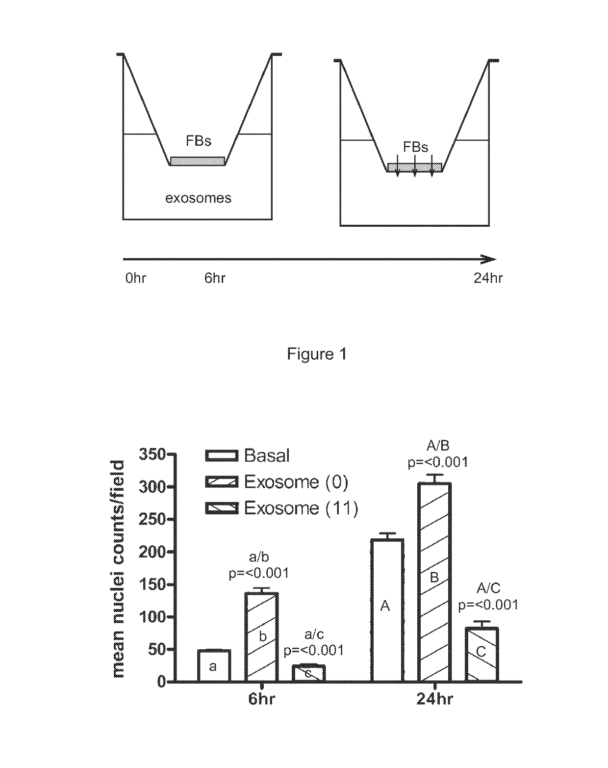
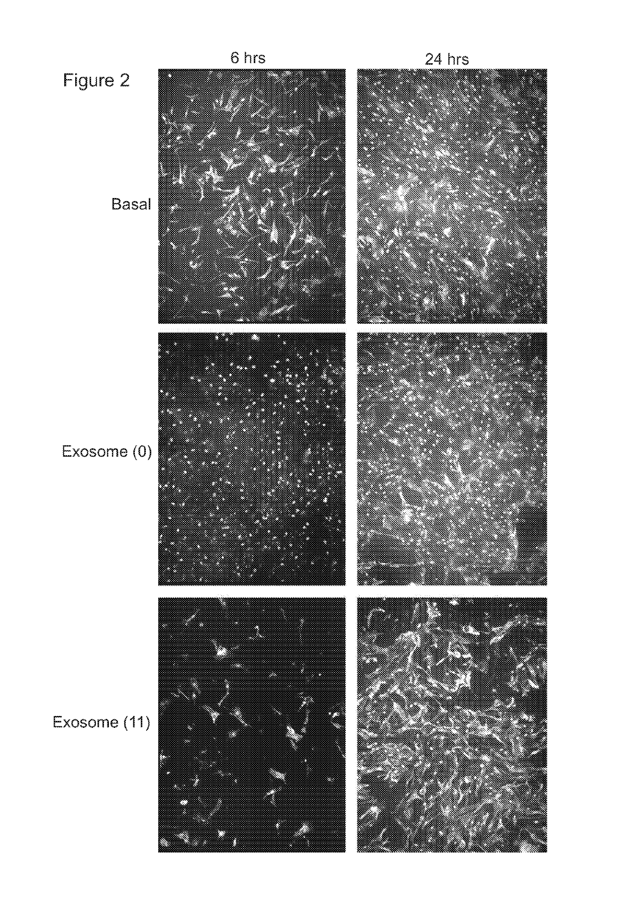
[0101]FIGS. 1 and 2 (Example 1) demonstrate that exosomes isolated from the conditioned medium of CTX0E03 cells that have been cultured for 11 weeks have the ability to inhibit fibroblast migration in a transmembrane assay model of cell migration. Accordingly, one biological function that microparticles of the invention may retain is the ability to inhibit migration of fibroblast or fibroblast-like cells, for example of normal human dermal fibroblasts (NHDF).

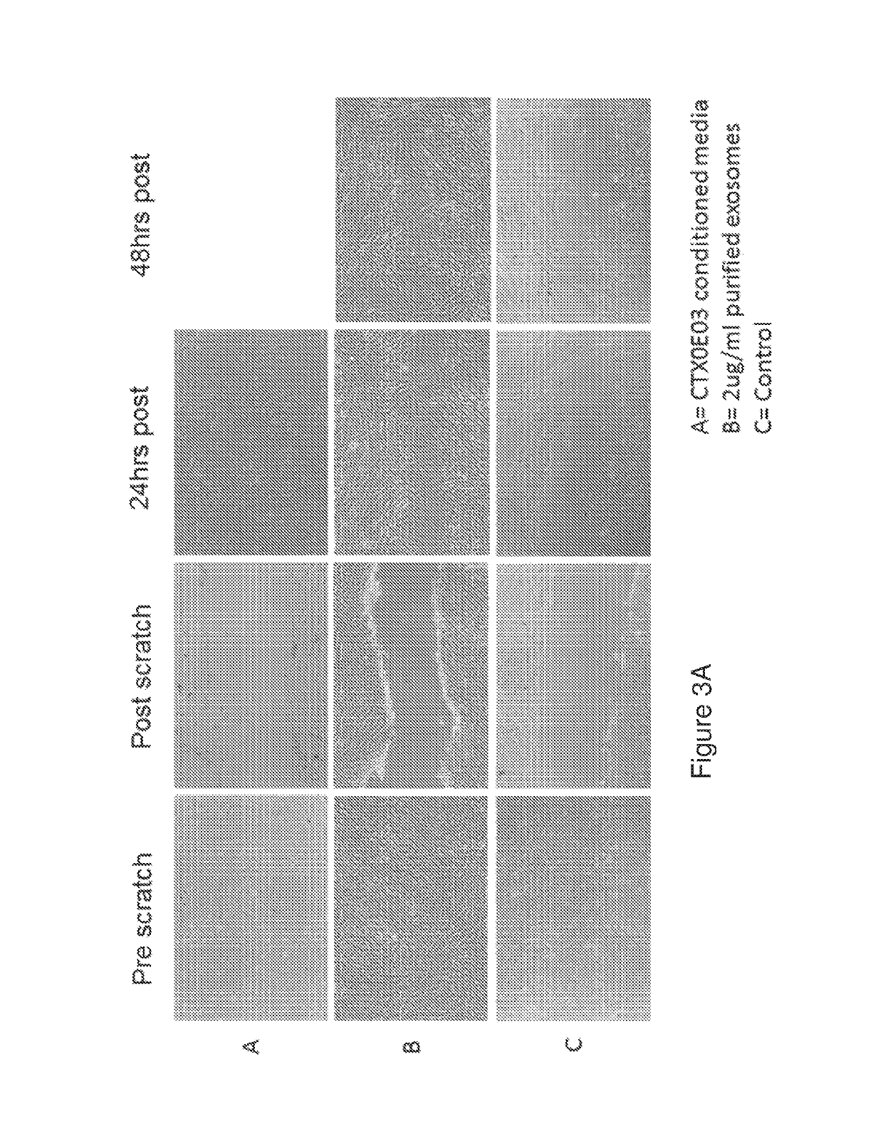
[0102]In contrast, exosomes isolated from the conditioned medium of CTX0E03 cells that have been cultured for 0-6 weeks promote cell migration as determined using a scratch / wound closure assay. Examples 1 and 2, Table 2 and FIGS. 1-3 demonstrate that exosomes isolated from the conditioned medium of CTX0E03 cells that have been cultured for 0-6 weeks retain the ability to close a wound in a “scratch” model of wound healing. The results in FIG. 3A show that the migration activity of normal human dermal fibroblasts (NHDF) cultured ...

Example

[0108]An alternative transmembrane assay was used in Example 6, as shown in FIG. 22. Here, glioblastoma cells are seeded on one side of the porous filter membrane. These cells are either seeded together with 20 μg / ml microparticles (FIGS. 22A and C) or have been pre-treated with 10 μg / ml microparticles for 24 hours (FIG. 22B). Medium containing a chemoattractant is placed on the opposing (lower) side. In Example 6, the chemoattractant is FBS. After an incubation period (e.g. 6-24 hours), the membrane is fixed and stained to reveal migrated cells (e.g. cell nuclei). The number of cells which have migrated through the pores of the membrane is again counted microscopically.

[0109]Cell migration is calculated as the number of cells that have migrated through the pores of the membrane in relation to basal conditions (without the microparticles). Inhibition of cell migration in this assay may typically be defined as a decrease in the number of cells that have migrated through the membrane,...

Example

[0114]The proteomic analysis in Example 18 indicates that neural stem cell exosomes comprise biological functions associated with the production, packaging, function and degradation of genetic material. Accordingly, in one embodiment, exosomes of the invention retain these functions, typically one or more of RNA polymerase function, RNA degradation function, ribosome function and spliceosome function.

[0115]The microparticle obtained from the neural stem cell has a diameter of 1000 nm or less. Typically, the microparticle of the invention will have a diameter of 200 nm or less, for example 100 nm or less. As noted in Table 1 above, microvesicles have a diameter of 100 nm to 1000 nm. Exosomes are typically defined as having a diameter of 30-100 nm, but more recent studies confirm that exosomes can also have a diameter between 100 nm and 200 nm, (e.g. Katsuda et al, Proteomics 2013 and Katsuda et al, Scientific Reports 2013). Accordingly, exosomes typically have a diameter between 30 n...


PUM

PropertyMeasurementUnit
Timeaaaaaaaaaa
Timeaaaaaaaaaa
Densityaaaaaaaaaa

Abstract

This invention relates to stem cell microparticles and miRNA isolated from these microparticles, their use and production thereof, in particular neural stem cell microparticles and their use in therapy of cancer, typically a nestin-positive cancer. The cancer may be glioma, melanoma, breast cancer, pancreatic cancer or prostate cancer. The stem cell microparticle is typically an exosome or microvesicle and may be derived from a neural stem cell line. The neural stem cell line may be a conditionally-immortalized stem cell line such as CTX0E03 (deposited at the ECACC with Accession No. 04091601).

Description

CROSS-REFERENCE TO RELATED APPLICATIONS[0001]This application is a national stage application under 35 U.S.C. § 371 of International Application No. PCT / GB2014 / 053044, filed Oct. 9, 2014, which in turn claims priority to United Kingdom Patent Application No. 1317887.6, filed Oct. 9, 2013, and International Application No. PCT / GB / 2014 / 052509, filed Aug. 14, 2014, the content of each of which is hereby incorporated by reference into this application in its entirety.FIELD OF THE INVENTION[0002]This invention relates to stem cell microparticles and miRNA isolated from these microparticles, their use and production thereof, in particular neural stem cell microparticles and their use in therapy.BACKGROUND OF THE INVENTION[0003]Stem cells have the ability to self-renew and to differentiate into functionally different cell types. They have the potential to be a powerful therapeutic tool, for example in the growing field of Regenerative Medicine, in particular regenerative therapy requiring ...

Claims


Application Information

Patent Timeline
IPC IPC(8): C12N15/113C12N5/0797A61K35/30A61K9/00
CPCA61K35/30C12N5/0623C12N15/113C12N2501/15C12N2501/24C12N2501/25C12N2502/088C12N2310/141A61K9/0019A61P35/00A61P35/04
Inventor HICKS, CAROLINESINDEN, JOHNSTEVANATO, LARACORTELING, RANDOLPH
Owner RENEURON LTD
Why Eureka
Try Eureka