

Enzymatically activatable peptide-redox modulator conjugates and use thereof
a technology of peptide-redox modulator and conjugate, which is applied in the direction of peptides, drug compositions, peptides/protein ingredients, etc., can solve the problems of still suffering drawbacks and the unfulfilled need to develop novel approaches
- Summary
- Abstract
- Description
- Claims
- Application Information
AI Technical Summary
Benefits of technology
Problems solved by technology
Method used
Image

Examples
example 1
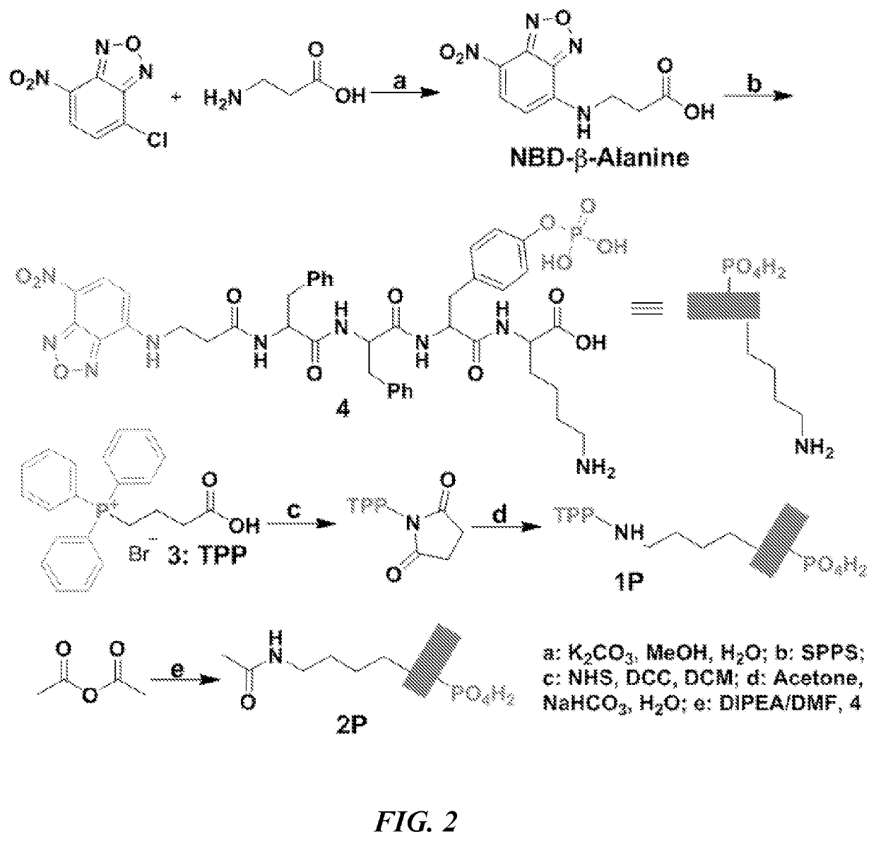
Design and Synthesis
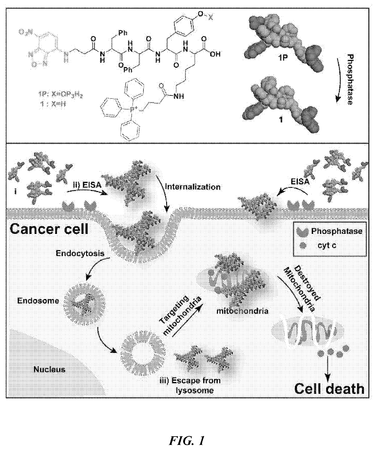
[0202]FIG. 1 shows the representative structure of the molecules designed for integrating EISA with mitochondria targeting. The molecules consist of four key features: a self-assembling backbone (i.e., a peptide containing D- or L-Phe-Phe-Tyr-Lys (FFYK)), an enzymatic trigger (i.e., tyrosine phosphate (pY) as a substrate of ALP), an environment-sensitive fluorophore (4-nitro-2,1,3-benzoxadiazole (NBD)-β-Ala), and a mitochondria targeting motif (i.e., TPP). FFYK was chosen because tyrosine provides a facile way to introduce the enzymatic triggers and FFY has acted as a motif for EISA (Gao et al., J. Am. Chem. Soc. 131:13576 (2009), which is hereby incorporated by reference in its entirety). NBD was used because NBD is a sensitive fluorophore for reporting molecular self-assembly in cellular milieu (Gao et al., Nat. Commun., 3:1033 (2012); Gao et al., Langmuir 29:15191 (2013); Zhou et al., J. Am. Chem. Soc. 137:10040 (2015); Gao et al., ACS Nano 7:9055 (2013), whic...
example 2
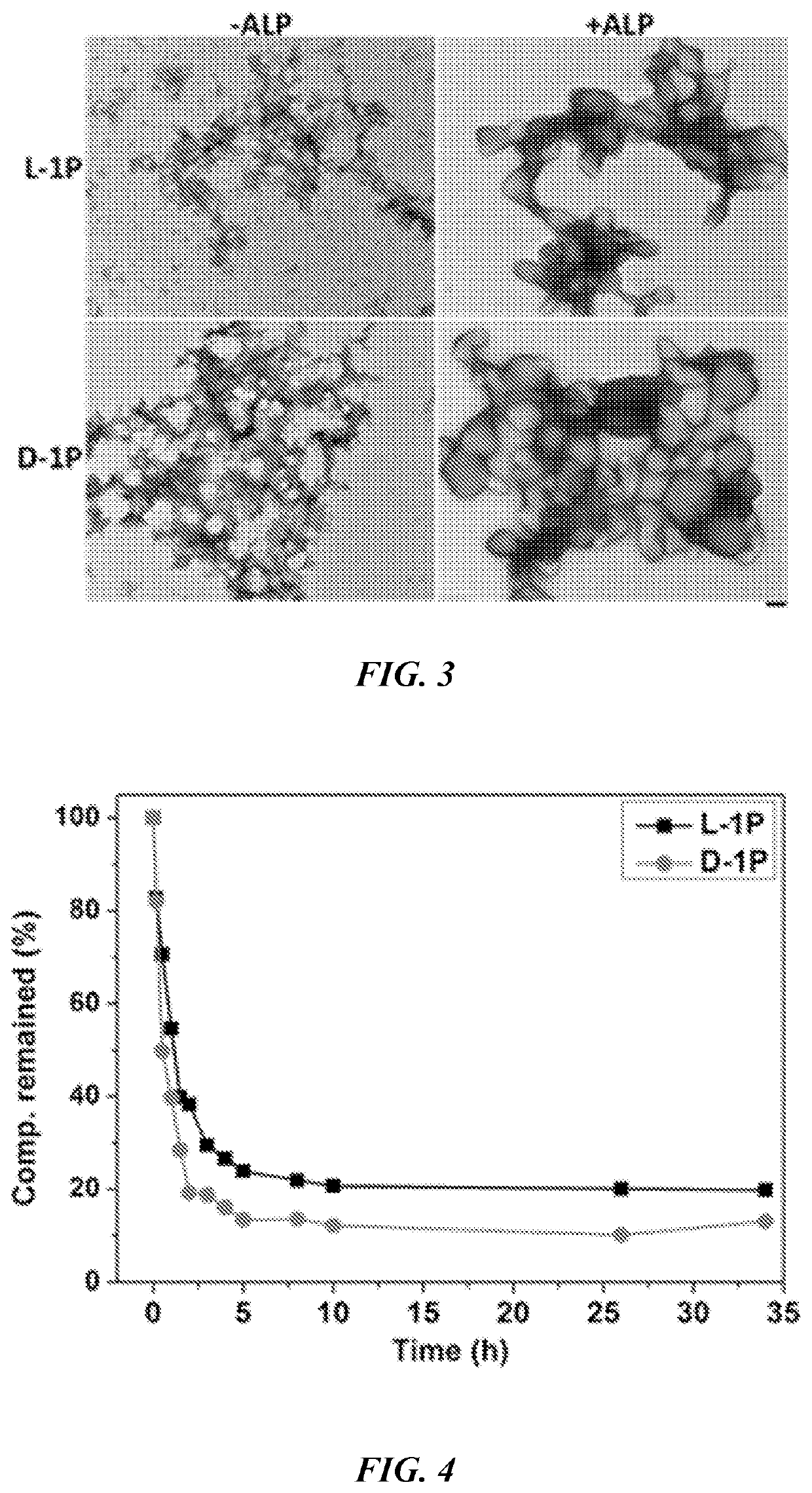
[0210]After obtaining all the precursors in Example 1, their behaviors were evaluated for EISA in vitro by using TEM and static light scattering (SLS) to examine the nanostructures formed before and after the addition of ALP into the solutions of the precursors. After drying from solution, L-1P (50 μM) shows many tiny nanoparticles with diameter of 5±2 nm, which tend to aggregate to result in irregular fibrous structures with diameter of 7±2 nm (FIG. 3), while at higher concentration, L-1P (100 μM) mainly forms irregular fibrous structure with few oligomers. As a contrast, D-1P (50 μM) forms slightly more regular fibrous structures with diameter of 8±2 nm, which then interact with each other to form dense 2D / 3D networks. Interestingly, D-1P (100 μM) forms more uniform nanoparticles with diameter of 25±2 nm. As revealed by the dephosphorylation experiment (FIG. 4), D-1P undergoes ALP catalyzed dephosphorylation slightly faster than L-1P does. The t1 / 2. was deter...
example 3
ity and Selectivity
[0212]To investigate the cellular response to all the precursors, an MTT (3-(4,5-dimethylthiazol-2-yl)-2,5-diphenyl tetrazolium bromide) assay (Gerlier et al., J. Immunol. Methods 94:57 (1986), which is hereby incorporated by reference in its entirety) was first used to examine the viability of human osteosarcoma cells (Saos2, which expresses high level ALP (Farley et al., Metabolism 40:664 (1991), which is hereby incorporated by reference in its entirety)) cultured with the precursors. As a control, the viability of normal human bone marrow stromal cells (H55) that express low level of ALP on cell surface was also examined (Zhou et al., Chem 1:246 (2016), which is hereby incorporated by reference in its entirety). As shown in FIG. 7, L-1P exhibits IC50 of 61±2 μM (76.1±2.5 μg / mL, 48 h) against Saos2 cells in a dosage dependent manner. D-1P exhibits IC50 of 46±2 μM (57.4±2.5 μg / mL, 48 h), lower than the IC50 of L-1P. In the presence of exogenous ALP, L-1P (or D-1P...
PUM
| Property | Measurement | Unit |
|---|---|---|
| Molar density | aaaaa | aaaaa |
| Molar density | aaaaa | aaaaa |
| Concentration | aaaaa | aaaaa |
Abstract
Description
Claims
Application Information