Nanocomposite Ionic-Covalent Entanglement Reinforcement Mechanism and Hydrogel

a technology of ionic-covalent entanglement and nanocomposite, which is applied in the direction of additive manufacturing processes, prostheses, manufacturing tools, etc., can solve the problems of poor biodegradability, weaker and harder characterization, and the material has little to no cell-material interaction, so as to achieve the effect of superimposing, dispersing energy and maintaining elasticity

Active Publication Date: 2020-03-05
TEXAS A&M UNIVERSITY
View PDF0 Cites 6 Cited by
  • Summary
  • Abstract
  • Description
  • Claims
  • Application Information

AI Technical Summary

Benefits of technology

[0012]The strengthening mechanisms captured in the present compositions, structures, and constructs with the disclosed bioinks have superior adaptability and utility for the techniques and biotechnologies disclosed here.
[0043]Single component hydrogel networks (like MA-kCa) collapse under stress as cracks concentrate stress and propagate throughout the network. In contrast, ICE networks become tougher by transferring stress between the separate networks, preventing any one zone from being overloaded. The brittle ionic bonds dissipate energy by reversibly breaking their crosslinks, while the flexible covalent network maintains elasticity and prevents crack propagation. Nanosilicates also toughen the network through charge interactions that stabilize the polymer chains, resisting deformation.

Problems solved by technology

[10] However, while polysaccharides and synthetic polymers are easily characterized and crosslinked, these materials have little-to-no cell-material interaction and poor biodegradability.
Natural protein hydrogels have superior bioactivity and are enzymatically degradable, but may be weaker and harder to characterize.
Despite existing developments, most bioinks are still poorly suited to printing 3-D structures.
Sufficient printability and strength in a bioink are problems that create challenges in the utilization of 3-D printing in biological / physiological applications for a number of reasons.
In particular, conventional bioinks upon extrusion have inferior structural strength when extruded to form layers, and the bioink layers are observed to quickly spread when placed in a multi-bioink layer construct from the weight of additional layers.
This problem precludes the formation of clinically useful bioprinted structures that require a height of more than about half a centimeter.

Method used


Image

  • Nanocomposite Ionic-Covalent Entanglement Reinforcement Mechanism and Hydrogel
  • Nanocomposite Ionic-Covalent Entanglement Reinforcement Mechanism and Hydrogel
  • Nanocomposite Ionic-Covalent Entanglement Reinforcement Mechanism and Hydrogel

Examples


example 1

Materials and Methods—Synthesis and In Vivo Integration and Biodegradability

[0058]The present example describes the bioink composition and synthesis thereof, as well as the use of the bioink in the creation of a multi-layer, 3-D, bioink construct / structure suitable for in vivo and / or clinical use.

[0059]Bioink Composition

[0060]The NICE bioink was made of 10% w / v (80% methacrylated) gelatin methacrylate, 1% w / v kappa carrageenan (KCa), 2% w / v Laponite XLG, and 0.25% w / v Irgacure 2959 2-Hydroxy-4′-(2-hydroxyethyoxy)-2-methylpropriophenone as a photoinitiator. The nanosilicates (Laponite XLG) were sourced from BYK Additives Inc. The porcine gelatin (gel strength 300, Type A) was obtained from Sigma. Irgacure 2959 and Methacrylic Anhydride were both obtained from Aldrich.

[0061]Bioink Synthesis

[0062]Gelatin methacrylate (GelMa) was synthesized by dissolving 10 g of gelatin in 100 mL 1× phosphate buffered saline (PBS), then heating for 1 hour at 60° C. After dissolution, 8 mL of methacryli...

example 2

Nanocomposite Reinforcement

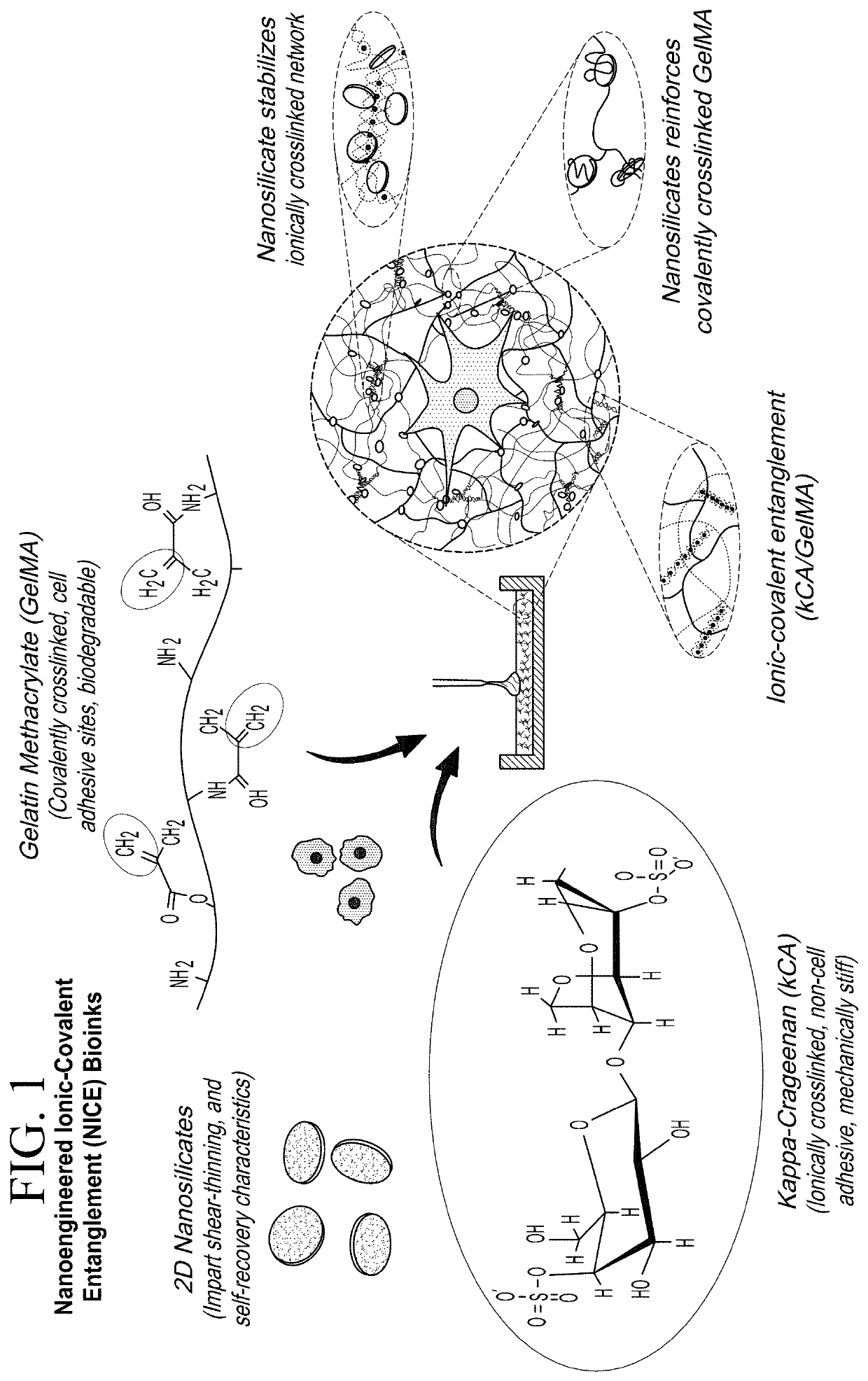
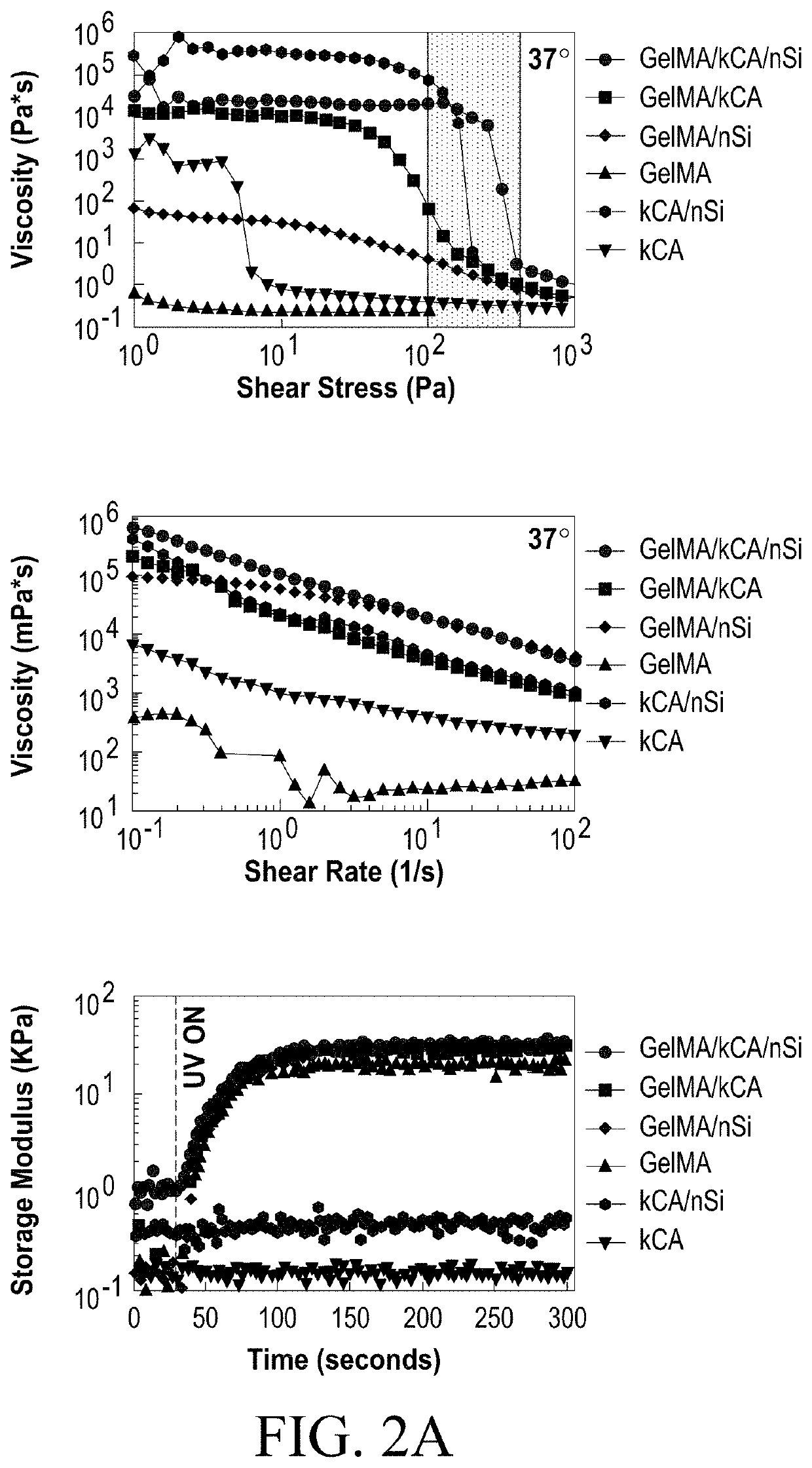
[0090]The nanocomposite reinforcement was accomplished by inclusion of 2% (w / w) Laponite XLG nanoparticles. Laponite nanoparticles have negatively charged faces and a positively charged rim, which allow Laponite to form reversible electrostatic interactions with the polymer backbones of hydrogels, effectively acting as a weak secondary crosslinker. This interaction can improve stiffness, elasticity, adhesiveness, viscoelastic modulus, and cell adhesion in some hydrogels, and imbue hydrogel solutions with complex shear thinning and bingham plastic behavior (FIG. 1). In the NICE bioink, Laponite forms reversible bonds with both gelMa and k-carrageenan polymers, strengthening the bioink before and after crosslinking, and improving its viscoelastic properties (FIG. 1).[17, 25-31]

[0091]Ionic covalent entanglement (ICE) networks are composed of two independent-but-entangled polymer networks that are not crosslinked to each other thanks to distinct crosslinking m...

example 3

Blood Vessel 3-D Bioprinting

[0094]The printability of the NICE bioink was evaluated through the present studies to illustrate the reproducibility and objectiveness of the material for facilitating direct comparisons with other bioinks. As previously noted, “printability” is defined as a bioink's ability to print high aspect ratio structures at animal (human)-relevant scales and extrude the intended scaffold architecture smoothly and with high fidelity.

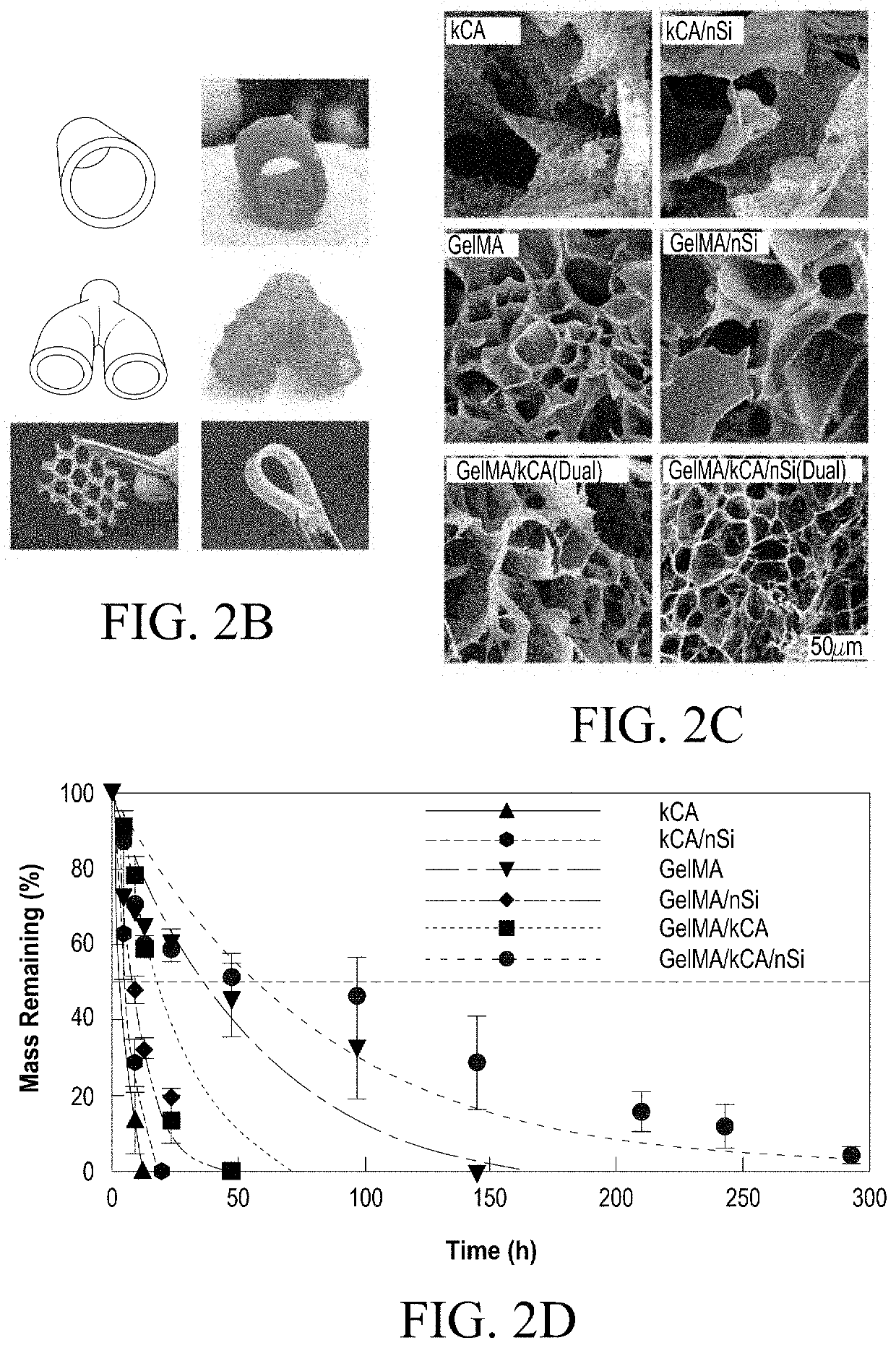
[0095]Printability of the present methods and materials is demonstrated here with a cylindrical print test of a “blood vessel” shape 1 cm in diameter with 1 mm thick walls (FIG. 4B), approximating the scale of a human blood vessel. This construct can be used as a standard to quantify aspect ratio, maximum construct height, and bioink spreading to allow direct comparison to other bioinks.

[0096]Minimizing bioink spreading is necessary for printing high fidelity structures, and was evaluated using the cylindrical print test to a height of...


PUM

PropertyMeasurementUnit
aspect ratioaaaaaaaaaa
widthaaaaaaaaaa
aspect ratioaaaaaaaaaa

Abstract

A biodegradable and biocompatible three dimensional construct comprising a combination of a nano silicate (e.g., laponite) and two different polymers, the two polymers each individually providing at least one covalently linked polymer chain and at least one ionically linked polymer chain, the polymeric chains forming a dual strengthening intertwined polymeric system. The constructs demonstrate improved mechanical and strength properties, while the bioinks provide a material having superior printability characteristics suitable for printing a three dimensional biodegradable construct having an aspect ratio of greater than 2.0. The bioink may also comprise cells or combinations of cells. Methods of using the constructs and bioinks for wound healing preparations and tissue regeneration are also provided.

Description

STATEMENT REGARDING FEDERALLY SPONSORED RESEARCH OR DEVELOPMENT[0001]This invention was made with government support under NIH-RO1AR066033-01, NSF-CBET-1264848 and HRD-1406755. The government has certain rights to the invention.TECHNICAL FIELD[0002]This invention relates to the field of biologically compatible materials such as bioinks, and the creation and uses of bioinks in 3-D printing for tissue fabrication and repair.BACKGROUND ART[0003]3-D bioprinting is emerging as a promising method for rapidly fabricating human biomimetic tissue constructs using cell-containing hydrogels, or bioinks, that are then crosslinked to form a viscoelastic matrix for the encapsulated cells. 3-D bioprinting innovation is driven by the clinical need for creating healthy and functional tissues for integration into a patient's body. However, the field of 3-D bioprinting remains in need of available and more suitable bioinks that are capable of printing structures having sufficient height greater than a...

Claims


Application Information

Patent Timeline
Patent Type & Authority Applications(United States)
IPC IPC(8): C09D11/101B29C64/118B33Y10/00B33Y70/00B33Y80/00C09D11/03C09D11/04C09D11/107A61L26/00A61L27/02A61L27/50A61L27/52A61L27/54A61L27/58A61L27/22A61L27/16A61L27/26
CPCA61L27/58B29K2105/162A61L27/26A61L2400/06A61L27/025A61L27/52A61L26/0066A61L26/0052A61L27/507A61L26/0038C09D11/03A61L2400/04A61L26/0014A61L26/008B29K2509/00A61L27/54B29K2995/006A61L26/0004A61L26/0057C09D11/04A61L2400/12B33Y10/00A61L27/222A61L2300/412A61L26/0023C09D11/107A61L26/009C09D11/101B29K2995/0056A61L27/16B33Y70/00A61L2300/418B33Y80/00B29C64/118C09D11/02B29C64/106C09D11/037C09D11/08C09D11/10C09D11/14B33Y70/10
Inventor GAHARWAR, AKHILESH K.CHIMENE, DAVID
Owner TEXAS A&M UNIVERSITY
Why Eureka
Try Eureka