A method for characterizing melanocytic lesions

a melanocytic and lesions technology, applied in the field of melanocytic lesions characterization, can solve the problems of inability to distinguish between subtypes/variants of a disease, inability to discriminate between malignant and benign lesions, time-consuming and laborious, etc., to simplify a time-consuming and hence expensive procedure, and facilitate diagnosis

Inactive Publication Date: 2020-03-05
FRIEDRICH ALEXANDER UNIV ERLANGEN NURNBERG
View PDF0 Cites 1 Cited by
  • Summary
  • Abstract
  • Description
  • Claims
  • Application Information

AI Technical Summary

Benefits of technology

[0011]With this invention, including a list of specific tissue markers (n=7-10), this time-consuming diagnostic procedure is standardized, easy to diagnose by less experienced experts and suitable for automated staining and scanning platforms in the new age of digital pathology. The present invention allows the early and precise diagnosis of acute and malignant skin diseases that involve melanocytes, different clinical stages and diseases predisposing conditions by naked eye or s

Problems solved by technology

In addition, it is often unable to discriminate between subtypes/variants of a disease, a requirement for personalized medicine.
However, until today it remains merely in a supportive role usually restricted to stains with one antibody because of

Method used


Image

  • A method for characterizing melanocytic lesions
  • A method for characterizing melanocytic lesions
  • A method for characterizing melanocytic lesions

Examples


example

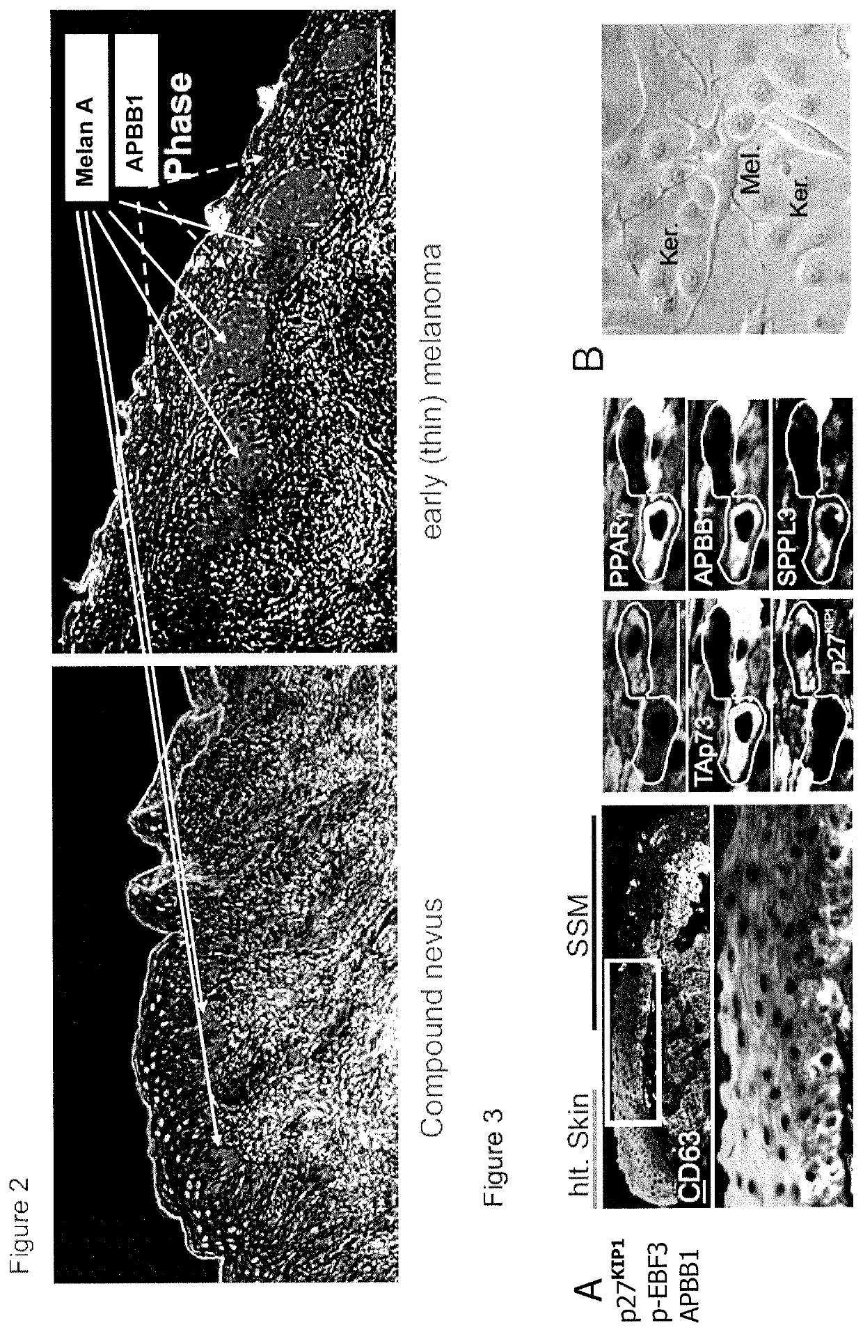
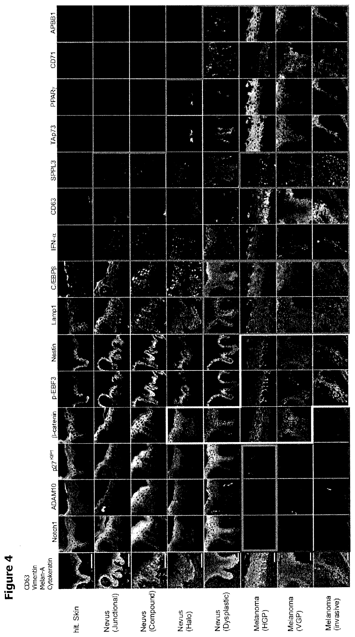
[0073]Using a systemic approach with the multi-epitope-ligand-cartography (MELC)-technology, we analyzed protein expression profiles (PEP) in nevi and BRAFV600E+ superficial spreading melanomas (SSM) for key transformation events.

[0074]To obtain antibodies applicable in the MELC-technology, 814 randomly selected hybridoma supernatants from the antibody production facility of the Helmholtz-Centre in Munich, and 173 commercially available antibodies were subjected to a screening algorithm to obtain those antibodies giving a specific staining in tissue (epidermis and dermis) for melanoma cells (n=57). We reasoned that key factors of the transformation process would appear in melanomas but not in nevi. We also selected antibodies that were specific for melanoma-associated keratinocytes (n=7) or for melanomas and keratinocytes (n=12). Single tissue sections of 6 BRAFV600E+ SSM, 6 junctional and compound nevi (3 / 3), and 6 samples of healthy skin were stained by the whole antibody set. For...


PUM

PropertyMeasurementUnit
Levelaaaaaaaaaa

Abstract

The present disclosure pertains to the identification of novel biomarkers for the characterization of melanocytic lesions as can be found in skin diseases such as melanomas and psoriasis. In particular, the present invention is directed to a method for characterizing melanocytic lesions and their use in an automated or semi-automated device for the diagnosis and/or monitoring of benign, pre-malignant, and malignant melanocytic lesions, as further defined in the claims.

Description

[0001]The present disclosure pertains to the identification of novel biomarkers for the characterization of melanocytic lesions as can be found in skin diseases such as melanomas and psoriasis. In particular, the present invention is directed to a method for characterizing melanocytic lesions and their use in an automated or semi-automated device for the diagnosis and / or monitoring of benign, pre-malignant, and malignant melanocytic lesions, as further defined in the claims.BACKGROUND OF THE INVENTION[0002]Human tissue is the most important source of biomarkers in health and disease, yet its analysis relies largely on classical histology which in essence is a hundred years old. At present histological diagnosis is still the gold standard in tissue analysis (pathology) supporting disease management in a crucial function, confirming or correcting the diagnosis when clinical appearance is inconclusive. In particular for tumour diagnosis and tumour classification (malignant / benign), his...

Claims


Application Information

Patent Timeline
IPC IPC(8): G01N33/574G16B40/10G01N35/00G16H50/30
CPCG01N33/5743G16H50/30G01N2035/00841G16B40/10G01N35/00584C12Q1/6886
Inventor BAUR, ANDREASOSTALECKI, CHRISTANSCHULER, GEROLD
Owner FRIEDRICH ALEXANDER UNIV ERLANGEN NURNBERG
Why Eureka
Try Eureka