Visualization and manipulation of results from a device-to-image registration algorithm

a technology of image registration and visualization, applied in the field of optical imaging, can solve the problems of a lot of variables, a lot of planning for ablation, and rarely good diagnostic imaging, and achieve the effect of optimizing the registration

Active Publication Date: 2020-04-23
CANON USA
View PDF4 Cites 5 Cited by
  • Summary
  • Abstract
  • Description
  • Claims
  • Application Information

AI Technical Summary

Benefits of technology

[0019]In at least one or more embodiments, automatic multi-planar reconstruction of an oblique fiducial slice may be provided. Upon user selection of a fiducial, the oblique slice may be automatically reconstructed provided a third dimension (3D) of registration. The oblique fiducial slice reconstruction may present all imaged fiducials of the device with overlaid representation of the virtual device model slice intersections as well as a representation of the detected fiducial locations. The oblique slice may allow the user to assess the virtual model matching to detected fiducial locations in the third dimension of the image. User manipulation of the detected fiducial locations, such as, but not limited to, adding, moving, and / or deleting fiducials, may be possible on this oblique fiducial slice reconstruction which drives a re-registration of the virtual model with the fiducial locations.
[0020]In at least one or more embodiments, on the fly (e.g., when needed, in real time, manual, automatic, during a medical procedure, while in progress, etc.) update(s) of template matching and registration error calculations based on user manipulation of detected fiducial locations (e.g., such as, but not limited to, adding, deleting, and / or moving fiducial locations) may be provided. As such, a user may receive, or be provided with, immediate feedback on how the user manipulation of detected fiducial locations affects the registration, and / or affects the matching of the virtual model and imaged fiducials (aka template matching), providing an intuitive method for the user to optimize the registration.
[0023]In one or more embodiments, the arc member comprises a guidance surface, wherein the guidance surface comprises one or more angular reference marks. The angular reference marks may be used to align with an indicator configured upon the arc member guidance surface to accurately situate the holder in the desired angular position (especially after the guide device is registered). In further embodiments, the medical guidance apparatus comprises a gap extending from the inner circumference of the frame to the outer circumference of the frame, to allow for detachment and / or reattachment of the medical guidance apparatus to the surface without interrupting the medical tool.
[0027]One or more other features discussed herein may reduce the risk of user error.
[0030]In accordance with at least another aspect of the present disclosure, the ablation probe or needle placement / guidance and / or visualization, manipulation and registration technique(s) discussed herein may be employed to reduce the cost of at least one of manufacture and maintenance of ablation planning and performance devices, systems and storage mediums by reducing or minimizing a number of components therein to cut down cost.

Problems solved by technology

However, diagnostic imaging is rarely good enough to plan with, so an IR may conduct initial imaging before developing / finalizing an action plan and starting an ablation procedure.
Ablation takes a lot of planning, and there are a lot of variables.
Even though medical procedures (e.g., ablation, biopsy, needle guidance, etc.) are very complex, the procedure that is currently performed by clinicians is predominantly done manually and iteratively, which is error prone and may increase the time required to perform, for example, an ablation (i.e., be inefficient).
However, it is often difficult for the user to quickly and easily visualize and manipulate the results of registration.
In 2D, marking a center position for an object may not be hard, even though many times it may not be accurate due to human visual and motor action inaccuracy / error.
However, a clinician using 2D slice view to figure out a center of a 3D volume which includes a stack of 2D slices may be difficult and error prone if the center of the volume is the target point, and the clinician may be tricked by image artifacts and / or human limitation in 3D reasoning.
In 3D, marking a center position is much harder because of the intricate limitation of visualization / rendering software.
Relying on clinicians' intuition, experience and visual understanding to define a target point is not optimal (for reliability, repeatability, traceability, etc.), particularly in 3D space.
When the lesion / tumor has a very complicated shape, defining an appropriate target is more or less an art, and it is difficult to achieve consistency.
Devices / hardware is also lacking to help clinicians aid in the insertion of multiple probes or needles and in the registration of such guidance devices, probes and / or needles during a procedure, such as, but not limited to ablation, biopsy, or other procedures.
It is very difficult to achieve these preset needle or probe configurations with this approach and thus an improved guidance, visualization and / or registration method is needed.
This not only increases the procedure time, but also wastes a lot of efforts in adjustment / making changes.
Of course, it is also likely having impact(s) on or causing possible damage to nearby structure and tissues.
Considering organ movement and deformation may make medical procedure planning and performance more complex, and may hamper interaction between clinicians and ablation planning and performance devices.
Such changes may also invalidate the planned insertion.
Respiratory gating, or asking patients to hold their breath, is time consuming monitoring techniques that may be used to assist with insertion.
However, such procedures do not guarantee success or efficiency.

Method used


Image

  • Visualization and manipulation of results from a device-to-image registration algorithm
  • Visualization and manipulation of results from a device-to-image registration algorithm
  • Visualization and manipulation of results from a device-to-image registration algorithm

Examples


Embodiment Construction

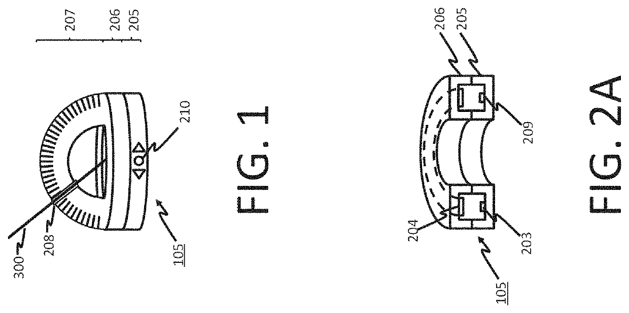
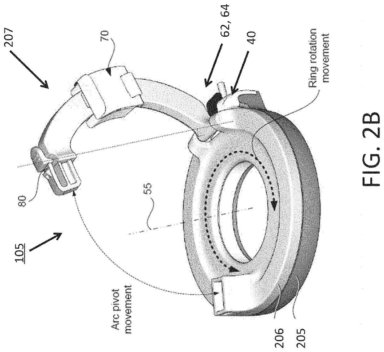
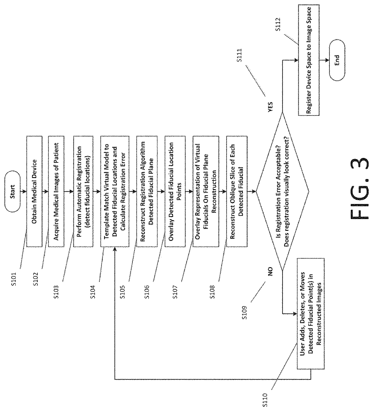
[0048]One or more devices, systems, methods and storage mediums for performing guidance for needles or probes and / or for visualization and manipulation of result(s) from device-to-image registration are disclosed herein. In one or more embodiments, the configurations, methods, apparatuses, systems and / or storage mediums may be combined to further enhance the effectiveness in guiding the needles or probes, including using one or more methods for visualization and manipulation of results from device-to-image registration to improve or optimize placement of the needle(s) and / or probe(s). Several embodiments of the methods, which may be carried out by the one or more embodiments of an apparatus, system and computer-readable storage medium, of the present disclosure are described diagrammatically and visually in FIGS. 1 through 14.

[0049]In accordance with at least one aspect of the present disclosure, at least one embodiment of a device for guiding needles or probes, and for performing v...


PUM


Abstract

One or more devices, systems, methods and storage mediums for performing medical procedure (e.g., needle guidance, ablation, biopsy, etc.) planning and / or performance, and / or for performing visualization and manipulation of registration result(s), are provided. Examples of applications for such devices, systems, methods and storage mediums include imaging, evaluating and diagnosing biological objects, such as, but not limited to, lesions and tumors, and such devices, systems, methods and storage mediums may be used for radiotherapy applications (e.g., to determine whether to place seed(s) for radiotherapy). The devices, systems, methods and storage mediums provided herein provide improved registration results by providing one or more of the following: automatic multi-planar reconstruction of a device fiducial plane, automatic multi-planar reconstruction of an oblique fiducial slice and on-the-fly update(s) of template matching and registration error calculations based on user manipulation of detected fiducial locations.

Description

CROSS-REFERENCE TO RELATED APPLICATION(S)[0001]This application relates, and claims priority, to U.S. Patent Application Ser. No. 62 / 747,913, filed Oct. 19, 2018, the entire disclosure of which is incorporated by reference herein in its entirety.FIELD OF THE INVENTION[0002]The present disclosure relates generally to the field of optical imaging, such as, but not limited to, in the field of interventional oncology, and more particularly to apparatuses, systems, methods and storage mediums for guidance of one or more medical instruments, such as needles used for minimally invasive puncture treatment, and for visualization and manipulation of device-to-image registration of such guidance apparatuses, systems, methods, and storage mediums. Examples of medical applications include imaging, evaluating and characterizing / identifying biological objects or tissue, such as, but not limited to, for identification, location and treatment of lesions / tumors, operation / procedure planning, simulati...

Claims


Application Information

Patent Timeline
Patent Type & Authority Applications(United States)
IPC IPC(8): A61B34/10A61B90/00A61B90/11A61B34/00A61B18/02
CPCA61B18/02A61B90/37A61B2018/0022A61B2018/0293A61B34/25A61B2018/00577A61B90/39A61B34/10A61B2018/0212A61B90/11A61B2034/107A61B2090/3937A61B2034/2059A61B2090/363A61B2090/3966A61B2034/104A61B2034/105A61B2090/3762A61B18/1477
Inventor DANIELS, BARRET
Owner CANON USA
Why Eureka
Try Eureka