Methods for scar reduction by converting scar fibroblasts into adipocytes with hair follicle-derived signals

a technology of scar fibroblasts and adipocytes, which is applied in the direction of drug compositions, skeletal/connective tissue cells, peptide/protein ingredients, etc., can solve the problems of scar fibroblasts almost universally recurring, and often affecting the appearance of scars, so as to reverse skin aging

Pending Publication Date: 2020-04-23
RGT UNIV OF CALIFORNIA +1
View PDF3 Cites 3 Cited by
  • Summary
  • Abstract
  • Description
  • Claims
  • Application Information

AI Technical Summary

Benefits of technology

[0005]The invention relates to pharmaceutical compositions and methods for treating wound healing pathologies, inhibiting or r...

Problems solved by technology

In some instances, these repair processes go awry and lead to wound healing pathologi...

Method used


Image

  • Methods for scar reduction by converting scar fibroblasts into adipocytes with hair follicle-derived signals
  • Methods for scar reduction by converting scar fibroblasts into adipocytes with hair follicle-derived signals
  • Methods for scar reduction by converting scar fibroblasts into adipocytes with hair follicle-derived signals

Examples


example 1

and Methods

[0121]Wounding Experiments.

[0122]All animal experiments were carried out in accordance to with the guidelines of the IACUC of the University of Pennsylvania and the University of California, Irvine. Full thickness 1.5×1.5 cm (2.25 cm2) excisional wounds were inflicted on the backs of three to eight week-old mice as described in M. Ito et al., Nature 447:316 (2007).

[0123]Phenotype Quantification Methodology.

[0124]Whole mount wounds were stained for Oil Red O and the number of regenerated hair follicles was counted from the skin surface, where each new hair is clearly visible. The number of new adipocytes was counted from the underside, where each adipocyte is clearly visible due to their very large size and prominent marking by Oil Red O dye. For each experiment, adipose tissue regeneration was quantified as the ratio of all new adipocytes / all new hair follicles (aka adipocyte / hair follicle index). The index values for all experiments, statistical significance and number o...

example 2

ion of Fat Cells from Mvofibroblasts During Wound Healing

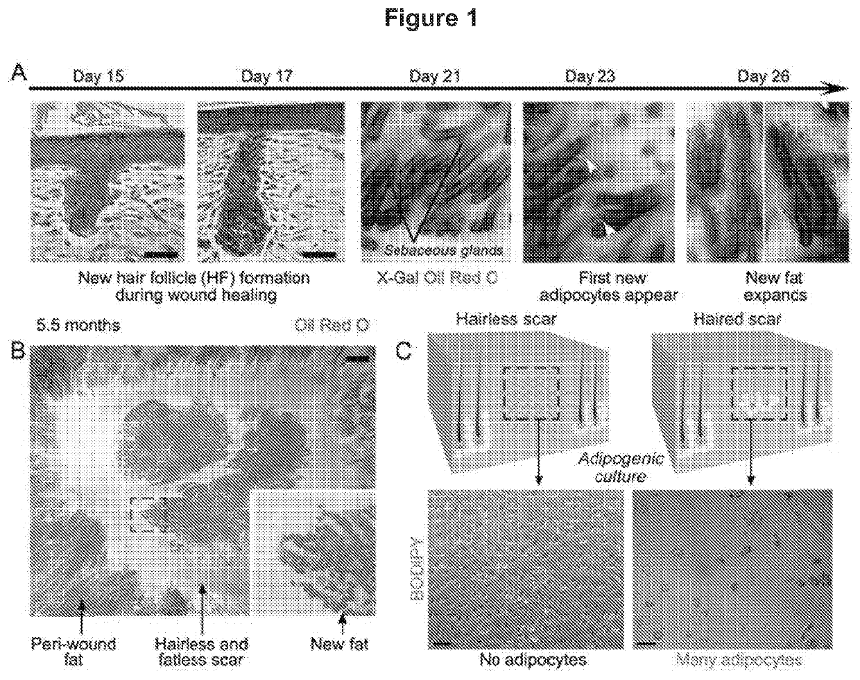
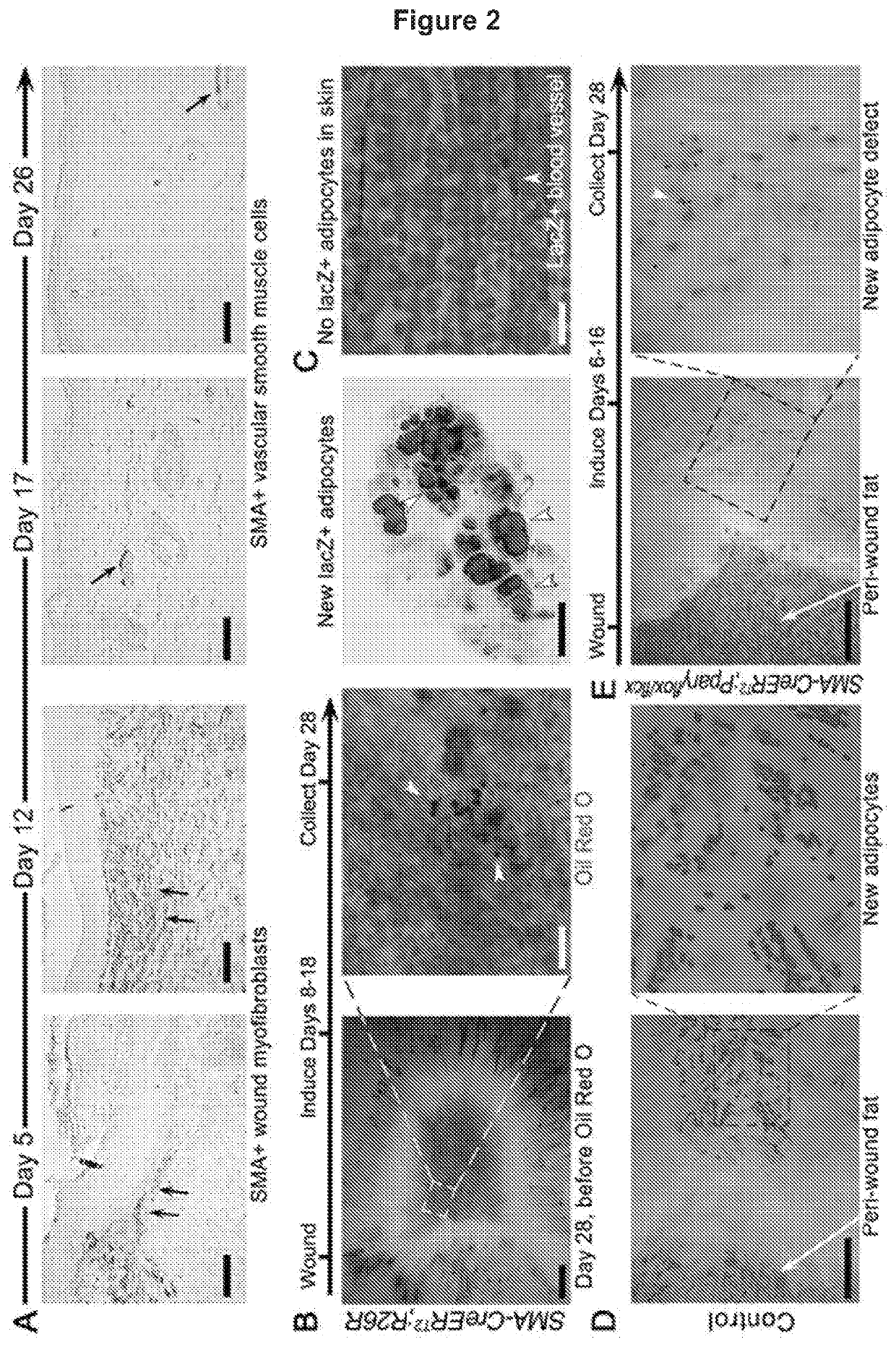
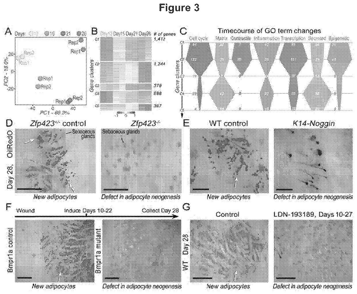
[0147]Although regeneration via the reprogramming of one cell lineage to another occurs in fish and amphibians, it is not observed in mammals. During wound healing adipocytes to regenerate from myofibroblasts, a cell type thought to be differentiated and non-adipogenic was discovered in mouse. Myofibroblast reprogramming required neogenic hair follicles, which triggered BMP signaling and then activation of adipocyte transcription factors expressed during development. Overexpression of the BMP antagonist, noggin, in hair follicles or deletion of the BMP receptor in myofibroblasts prevented adipocyte formation. Adipocytes formed from human keloid fibroblasts when treated with either BMP or when placed with human hair follicles in vitro. Thus, the myofibroblast is identified as a plastic cell type that may be manipulated to treat scars in humans.

[0148]Wound healing in adult humans and mice generally results in a scar with excess ...

example 3

l Lineage Tracing Results

[0161]Several non-myofibroblast mesenchymal cell types display SM22 / SMA activity in and around the wound bed. These include vascular smooth muscle cells (VSMCs) of the wound and peri-wound blood vessels, subcutaneous skeletal muscle panniculus carnosus and dermal papilla cells of new hair follicles. To definitively establish that myofibroblasts are the key mesenchymal adipogenic progenitors in the wound, a series of additional lineage tracing experiments were performed. First, we took advantage of the fact that not all smooth muscle contractile proteins are expressed in myofibroblasts. Smooth muscle myosin heavy chain (SMMHC aka Myhl 1) is expressed exclusively by true smooth muscle cells (18) and is distinctly absent from wound myofibroblasts. Lineage tracing in the wounds of SMMHC-CreERT2;R26R mice was performed using two induction protocols: 30 days before wounding to label pre-existing VSMCs of the cutaneous blood vessels and their progeny (n=4) (FIG. 13...


PUM


Abstract

This invention describes methods for treating wound healing pathologies, inhibiting or reversing fibrotic skin disorders, including but not limited to keloids, atrophic and hypertrophic scars, and reversing skin aging via generating new dermal adipocytes (DAs). Methods described herein comprise (a) grafting of human hair follicles or (b) direct delivery of conditioned media from in vitro cultured hair follicles, or (c) delivery of purified individual factors secreted by hair follicles, or (d) delivery of small molecule agonists that mimic hair derived signaling activities, or (e) delivery of small molecule antagonists that inhibit anti-adipogenic programs, or (f) delivery of small molecule agonists of pro-adipogenic programs to convert endogenous wound or scar skin fibroblasts into new DAs.

Description

CROSS-REFERENCE TO RELATED APPLICATIONS[0001]The subject Application is a U.S. national stage application under 37 U.S.C. 371 of PCT International Application PCT / US2018 / 012266, filed 3 Jan. 2018, which claims priority to U.S. Provisional Application No. 62 / 442,321, filed 4 Jan. 2017, the entire contents of which are hereby incorporated by reference in their entirety.GOVERNMENT INTEREST STATEMENT[0002]This invention was supported by Grant Numbers R01-AR055309, R01-AR067273, R01-AR069653, P30-AR057217, DK49210, GM055246, and T32-CA009054-37 from the NIH and DGE-1321846 from the NSF. The United States government has certain rights in the invention.FIELD OF THE INVENTION[0003]The invention relates to pharmaceutical compositions and methods for treating wound healing pathologies and aging, and regeneration of dermal adipocytes (DAs). Specifically, the invention relates to (a) canonical WNT pathway antagonists, (b) non-canonical WNT pathway ligands, (c) FGF pathway ligands, (d) BMP pathw...

Claims


Application Information

Patent Timeline
IPC IPC(8): A61K38/18A61K9/00A61K35/36A61P17/02A61K45/06
CPCA61K35/36A61P17/02A61K38/1875A61K9/0021A61K9/0014A61K45/06C12N5/0625C12N2501/155C12N2506/1307
Inventor COTSARELIS, GEORGEGUERRERO-JUAREZ, CHRISTIAN F.PLIKUS, MAKSIM
Owner RGT UNIV OF CALIFORNIA
Why Eureka
Try Eureka