Inhibition of vascular endothelial cell-mediated phagocytic processes for treatment of demyelinating conditions

a phagocytic process and demyelinating technology, applied in the field of demyelinating conditions inhibition of vascular endothelial cell-mediated phagocytic processes, can solve the problems of ineffective functional recovery of sci, high complexity of wound repair process,

Pending Publication Date: 2020-05-28
FLORIDA STATE UNIV RES FOUND INC
View PDF6 Cites 1 Cited by
  • Summary
  • Abstract
  • Description
  • Claims
  • Application Information

AI Technical Summary

Benefits of technology

[0008]It has been determined that microvessels and lining microvascular endovascular cells (ECs) act as amateur phagocytes to engulf myelin debris generated by disorders associated with demyelination. Mechanistically, the inventor determined that immunoglobulin G (IgG) opsonization of myelin debris is required for efficient uptake by microvascular ECs. The engulfed myelin debris is then delivered through the autophagy-lysosome pathway for intracellular degradation. Functionally, engulfment and autophagy-dependent processing of myelin debris by microvascular ECs contribute to three critical processes that are closely associated with demyelinating disorders: robust angiogenesis that results in excessive and abnormal microvessels, chronic inflammation, and endothelial-mediated fibrosis that most likely takes place through endothelial-to-mesenchymal transition (endoMT). Therefore, the inventor proposes that a benefit to patients can be obtained by intervening and interfering with the effects of myelin-ECs by targeting these particular processes (e.g., myelin debris uptake, autophagy and endoMT).
[0010]In some embodiments, the subject has a demyelinating condition at the time the agent is administered, and the agent is administered as therapy for the demyelinating condition. In other embodiments, the subject does not have a demyelinating condition at the time the agent is administered, and the agent is administered as prophylaxis to prevent or delay the onset of the demyelinating condition, including recurrence of a previous demyelinating condition. By treatment of a demyelinating condition in a subject, it is possible to reduce, prevent, or delay the onset of fibrosis, or a fibrotic condition, associated with vascular endothelial cell phagocytosis.

Problems solved by technology

The repair of wounds, for example, is a highly complex biological process.
Although tissue fibrosis, or scar formation, is a common response to damage in most organs of the body, and may be required for regaining tissue integrity, it is undesirable in many contexts.
For example, fibrosis resulting from nervous tissue is generally viewed as deleterious, as the fibrotic scar is inhibitory to regeneration and recovery of function.
Functional recovery of SCI is ineffective largely due to the failed or incomplete axon and myelin regeneration following SCI2, 3.
However, this cannot be the case for SCI because astrocytes are absent from the epicenter of injured spinal cords.

Method used


Image

  • Inhibition of vascular endothelial cell-mediated phagocytic processes for treatment of demyelinating conditions
  • Inhibition of vascular endothelial cell-mediated phagocytic processes for treatment of demyelinating conditions
  • Inhibition of vascular endothelial cell-mediated phagocytic processes for treatment of demyelinating conditions

Examples


example 1

els in the Demyelinating Spinal Cords Contain Myelin Debris

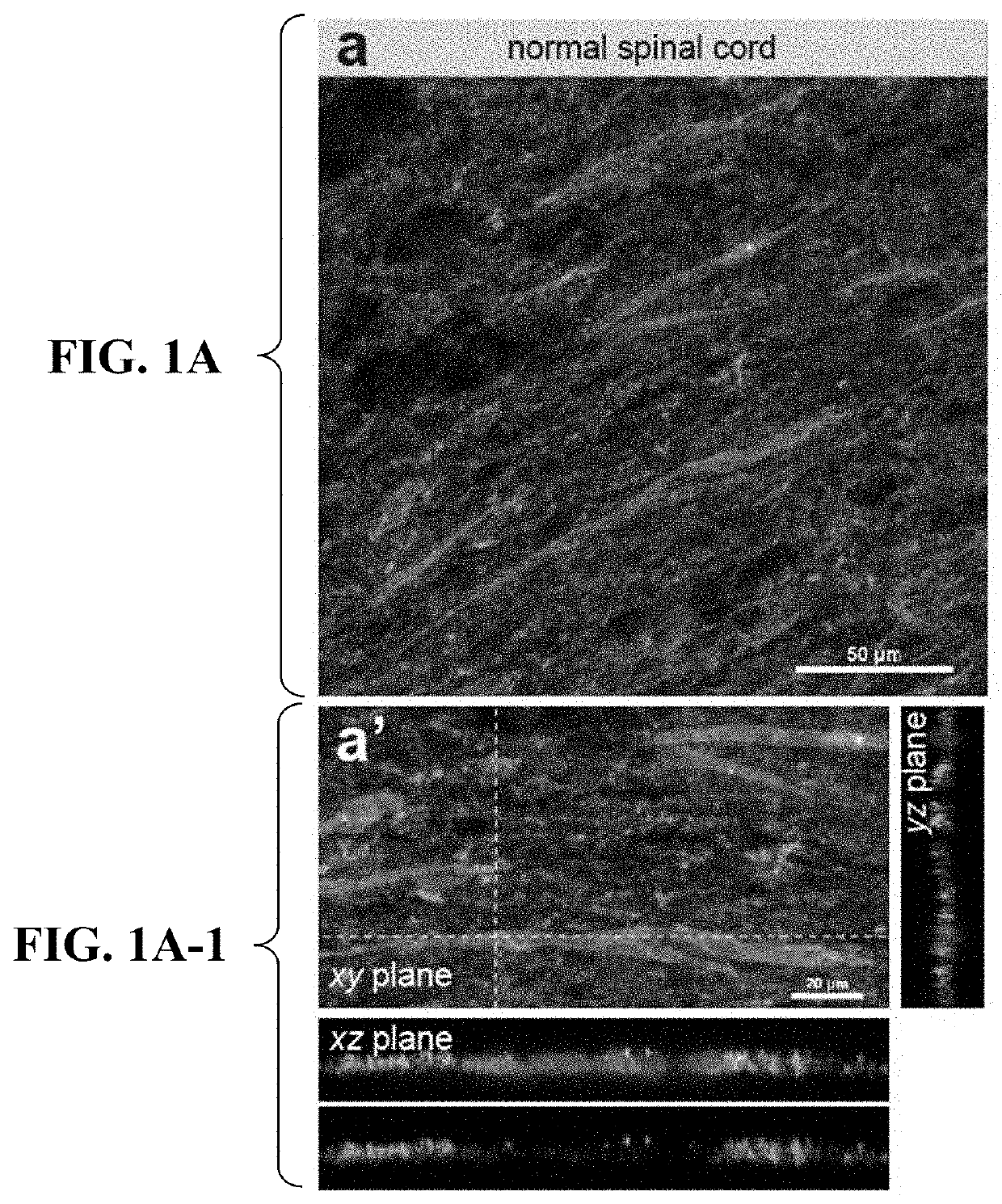
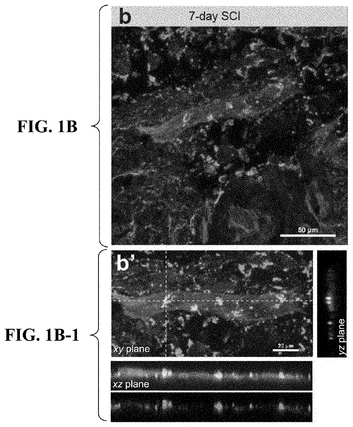
[0231]Microvessels in the lesion epicenter are lost during the first two days after SCI, whereas ECs proliferate and give rise to newly formed microvessels from 3 days after injury, restoring microvessel density to a normal level by 1 week after SCI15,16. We first examined whether these newly formed microvessels could engulf myelin debris. Myelin is intact in normal spinal cords and the uninjured spinal microvessels contain very little detectable myelin basic protein (MBP) (FIGS. 1A, 1A-1). By contrast, myelin debris, fragmented from myelin sheath following SCI, started to closely associate with newly formed microvessels in the lesion core as early as 3 to 5 days post SCI (FIG. 9), and became more apparent at 1 week after SCI (FIG. 1B). The x-z and y-z view of myelin debris distribution relative to microvessels revealed that myelin debris was indeed engulfed by microvessels (FIG. 1B-1, FIG. 9). Myelin debris-containing micro...

example 2

BMECs-Induced Microvessel-Like Structures Engulf Myelin Debris

[0233]Primary mouse brain microvascular endothelial cells (BMECs) grown on Matrigel as their substrate could form microvessel-like structures that mimics diverse aspects of microvessels20. After 72 hr incubation with microvessel-like tubules, CFSE (Carboxyfluorescein succinimidyl ester)-labeled myelin debris was seen as scattered puncta around or within the tubules (FIG. 1F). A closer inspection of the distribution of myelin debris from the confocal x-z view of a capillary lumen revealed the apparent dynamics of myelin debris entry (FIG. 1F-1). Some myelin fragments appeared to be close to but were not in direct contact with the capillary surface, as indicated by a lack of co-localization with endothelial marker CD31 (FIG. 1F-1). Other myelin fragments were in the process of entering, whereas still others had completely transited the luminal membrane, showing partial or full co-localization with CD31 (FIG. 1F-1). These da...

example 3

Engulfment of Myelin Debris by BMECs

[0234]We next investigated the kinetics and mechanisms of microvascular engulfment of myelin debris by using primary BMECs and a BMEC cell line bEnd.3. Both primary BMECs and bEnd.3 cells engulfed myelin debris in a time-dependent manner with predominant perinuclear distribution (FIGS. 2A, 2D). To quantitatively determine the efficiency and kinetics of myelin debris engulfment by BMECs, we used fluorescence-activated cell sorting (FACS) that counts the number of myelin-laden ECs (myelin-ECs), as well as a MBP ELISA assay to detect the intracellular MBP level. FACS results confirmed that primary BMECs were able to engulf myelin debris (FIG. 2B). MBP ELISA assay detected a significant amount of MBP in BMECs treated with myelin debris for 72 hr (FIG. 2C). The kinetics of myelin engulfment by bEnd.3 cells exhibited inefficient engulfment from 24-48 hr, efficient engulfment from 48-72 hr, and saturated engulfment from 72-96 hr (FIGS. 2E, 2F). As ECs ar...


PUM

PropertyMeasurementUnit
time pointsaaaaaaaaaa
molecular weightaaaaaaaaaa

Abstract

The present invention concerns a method for treating a demyelinating condition in a subject, by administering an agent to the subject that inhibits vascular endothelial cell phagocytosis. The method of the invention is useful in treating, for example, a demyelinating condition associated with an injury, such as a spinal cord injury or traumatic brain injury, as well as other demyelinating conditions, such as multiple sclerosis.

Description

CROSS-REFERENCE TO RELATED APPLICATION[0001]The present application claims the benefit of U.S. Provisional Application Ser. No. 62 / 770,968, filed Nov. 23, 2018, which is hereby incorporated by reference herein in its entirety, including any figures, tables, nucleic acid sequences, amino acid sequences, or drawings.GOVERNMENT SUPPORT[0002]This invention was made with government support under Grant No. DMS-0714589 and Grant No. DMS-1661727 awarded by the National Science Foundation. The government has certain rights in the invention.SEQUENCE LISTING[0003]The Sequence Listing for this application is labeled “2OT1984.TXT” which was created on Sep. 20, 2019 and is 8 KB. The entire contents of the sequence listing is incorporated herein by reference in its entirety.BACKGROUND OF THE INVENTION[0004]Fibrosis involves the overgrowth, hardening, and / or scarring of various tissues and is attributed to excess deposition of extracellular matrix (ECM) components, including collagen. Fibrosis can ...

Claims


Application Information

Patent Timeline
Patent Type & Authority Applications(United States)
IPC IPC(8): C12N15/113A61P25/28A61K9/00A61K38/47
CPCC12N15/113A61P25/28A61K9/0085A61K38/47C12N2320/32C12N2310/11C12N2310/14C12N2310/141C12Y302/01096C12N2310/20
Inventor REN, YI
Owner FLORIDA STATE UNIV RES FOUND INC
Why Eureka
Try Eureka