High-Throughput Platform for Bioprinting Tissue Modules

a tissue module and high-throughput technology, applied in biochemistry apparatus and processes, specific use bioreactors/fermenters, prosthesis, etc., can solve the problem of enhancing prevascularization in engineered tissues, and achieve the effect of rapid production of organized tissue precursors

Pending Publication Date: 2020-11-12
UNIV OF SOUTH FLORIDA
View PDF0 Cites 1 Cited by
  • Summary
  • Abstract
  • Description
  • Claims
  • Application Information

AI Technical Summary

Problems solved by technology

The most prevalent challenge encountered in the current techniques is the need to enhance prevascularization in the engineered tissues.

Method used


Image

  • High-Throughput Platform for Bioprinting Tissue Modules
  • High-Throughput Platform for Bioprinting Tissue Modules
  • High-Throughput Platform for Bioprinting Tissue Modules

Examples


example 1

ve Pressure Applied During Printing Affects Cell Viability

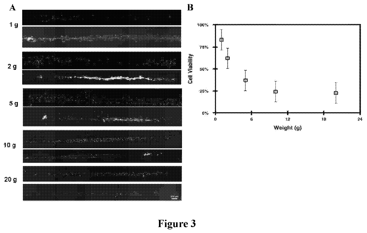
[0078]Viability of the printed tissue precursors was found to be predominantly correlated with the amounts of compressive pressure applied during printing. Cell survival was observed after 24 hours of printed tissues on polylysine coated target surfaces. To examine the adequate amounts of pressure necessary to print tissue modules, cells seeded on the shape-changing structures were subjected to pressures ranging from 0.71 to 14.5 psi and viability of the cells after transfer was analyzed. Calibration weights of 1-g, 2-g, 5-g, 10-g, and 20-g were used to vary the amounts of pressure. A LIVE / DEAD viability assay determined that the lowest amount of pressure applied resulted in the most viable cells (˜83%±3) of the (FIG. 3). The number of dead cells decreased as the amount of pressure applied was systematically reduced. A plot of the measured viable cells shows a power series regression model with an R-squared value of 95.9%.

example 2

Micro-contact Printing on Cell Adhesion Molecules Adsorbed Target Surfaces

[0079]Printing on target surfaces adsorbed with collagen type 1, fibronectin, and polylysine did not show any statistical difference in cell viability. Viability of printed tissue precursors was reduced by 11, 22, and 7% for collagen, fibronectin, and polylysine, respectively, in comparison to the control samples. The negative control consisted of cells printed on plain glass surface, and no cell transfer was observed to such surfaces while a positive control that included cells cultured on plain glass showed the highest number of viable cells. A single factor ANOVA test indicated no significant difference between the means (p>0.05). For subsequent printing, polylysine was selected as the CAM of choice for all target surfaces due to the ease of use and stability at ambient temperature.

example 3

issue Precursors Reveal Preserved Focal Adhesion Components

[0080]To assess cellular morphology and metabolic activity, printed cells were compared to cells cultured on a plain glass surface. An hour after printing, the actin stress fiber staining showed an elongated morphology compared to the rounded morphology seen in cells seeded on glass even after 2 hours. To examine the cell-matrix adhesions, the cell nucleus was stained and the alpha 5 integrin observed an hour and 2 hours after printing. The results reveal emergence of physical integrin clustering at an hour after printing and enhanced clustering after 2 hours of printing indicating rapid reformation and recapitulation of focal adhesion components, which is attributed to the preserved cell morphology compared to the control samples (FIG. 5).


PUM

PropertyMeasurementUnit
timeaaaaaaaaaa
weightsaaaaaaaaaa
weightsaaaaaaaaaa

Abstract

The invention pertains to methods and apparatuses for rapid production of organized tissue precursors with different sizes and geometry. An embodiment of the invention provides a method of producing 3D tissue structures, the method comprising: producing a plurality of patterned monolayers of cells and sequentially overlaying the plurality of patterned monolayers of cells to produce a stack of monolayers of cells. The patterns of cells in the plurality of monolayers can be designed in a manner which produces a tissue or a portion of a tissue when the plurality of monolayers is overlaid in to a stack. The invention also pertains to an apparatus for producing a 3D tissue module. The apparatus comprises: a three-dimensional positioning system, a contact stamp, a contact stamp storage container, a contact stamp holder, a contact stamp exchange unit, and a contact stamp actuator.

Description

CROSS-REFERENCE TO RELATED APPLICATION[0001]The present application claims the benefit of U.S. Provisional Application Ser. No. 62 / 237,843, filed Oct. 6, 2015, which is hereby incorporated by reference in its entirety, including any figures, tables, or drawings.[0002]This invention was made with government support under Grant Numbers DMR-0645574, DMR-1056475, and DGE-0638709 awarded by the National Science Foundation. The government has certain rights in the invention.FIELD OF THE INVENTION[0003]The invention provides methods and apparatuses for producing three-dimensional tissue structures.BACKGROUND OF INVENTION[0004]Engineering robust biomimetic 3D tissue structures is crucial to facilitate the repair of diseased tissues and organs. An ideal tissue construct should be able to recapitulate the functions of complex tissue micro architecture found in natural tissues. Construction of 3D tissues in vitro also creates an effective design model to systematically study a variety of cell ...

Claims


Application Information

Patent Timeline
Patent Type & Authority Applications(United States)
IPC IPC(8): C12N5/00C12M3/00B33Y10/00B33Y80/00
CPCB33Y80/00C12M21/08C12N5/0062B33Y10/00C12N2513/00A61L27/52A61L27/18A61L27/38A61L27/3886A61L27/50C08L83/04
Inventor CROSS, MICHAEL C.AKINTEWE, OLUKEMI O.DUPONT, SAMUEL JAMESELINENI, KRANTHI KUMARTOOMEY, RYAN G.GALLANT, NATHAN D.
Owner UNIV OF SOUTH FLORIDA
Why Eureka
Try Eureka