Assay for jc virus antibodies
a technology of jc virus and antibody, applied in the field of antibodies for jc virus, can solve the problems of insensitivity of methods, limited use, and seroprevalence ra
- Summary
- Abstract
- Description
- Claims
- Application Information
AI Technical Summary
Benefits of technology
Problems solved by technology
Method used
Image
Examples
example 1
and Purification of Highly Purified VP1 Particles
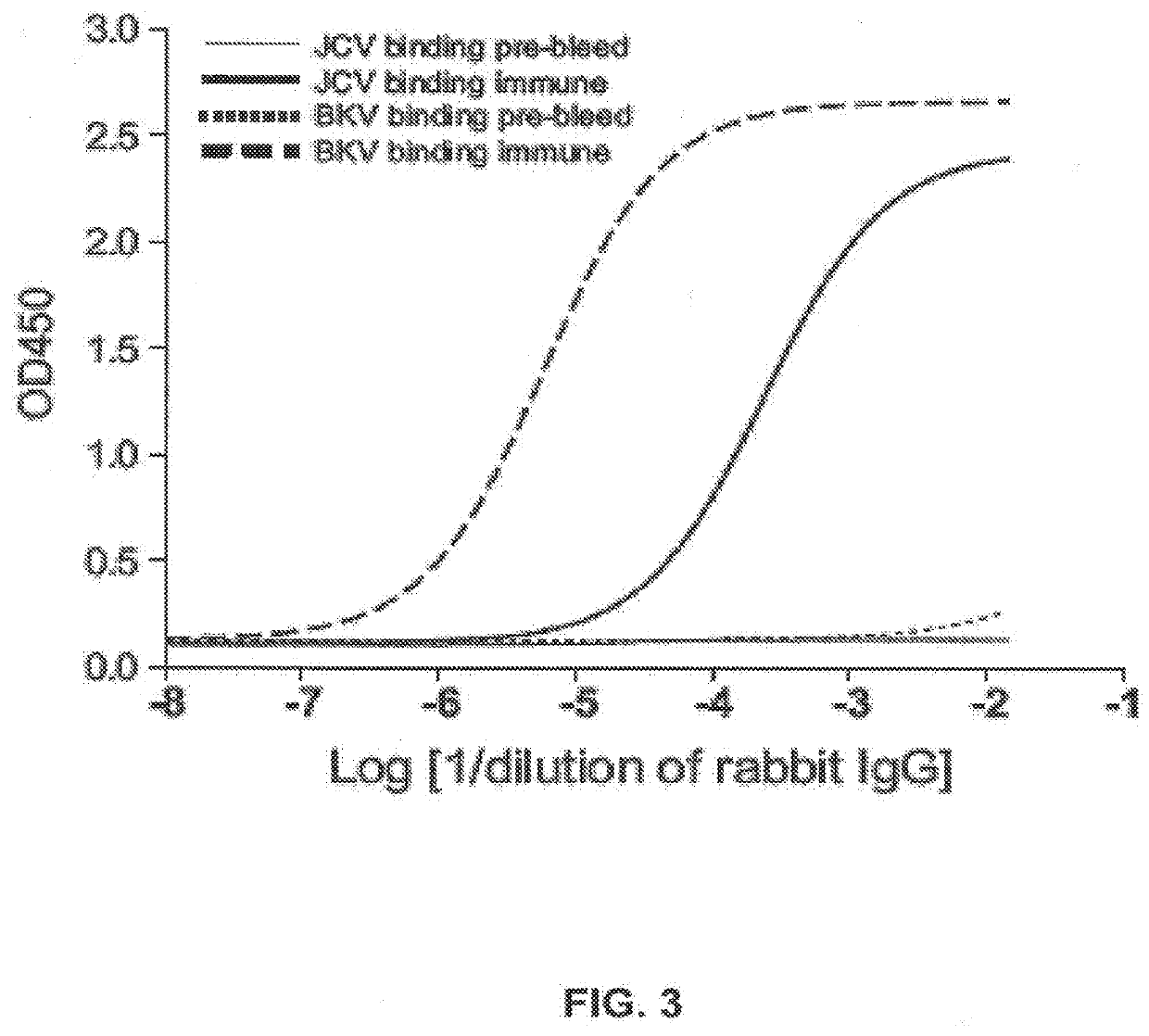
[0085]HPVLPs consisting of JCV or BKV capsid protein VP1 were produced in SF9 insect cells transfected with a recombinant baculovirus. In the case of JCV VP1 containing particles, recombinant baculovirus was transformed with a nucleic acid expressing VP1 from the Mad-1 strain of JCV. The recombinant VLP was harvested prior to cell lysis and was purified by differential ultracentrifugation, detergent washing and ultrafiltration.
[0086]Briefly, baculovirus infected cells were harvested about three days post infection by centrifugation at 3000×G and stored frozen until purification of HPVLPs. Purification was performed using about 100 grams of frozen cell pellets. Thawed cells were lysed in 500 ml of PBS supplemented with 0.1 mM CaCl2 (PBS-C). The cells were disrupted by passing the cell suspension twice through a Microfluidics Microfluidizer®. Cell debris was removed by pelleting at 8000×G for 15 minutes. The supernatant volume was adjus...
example 2
ibody Assay
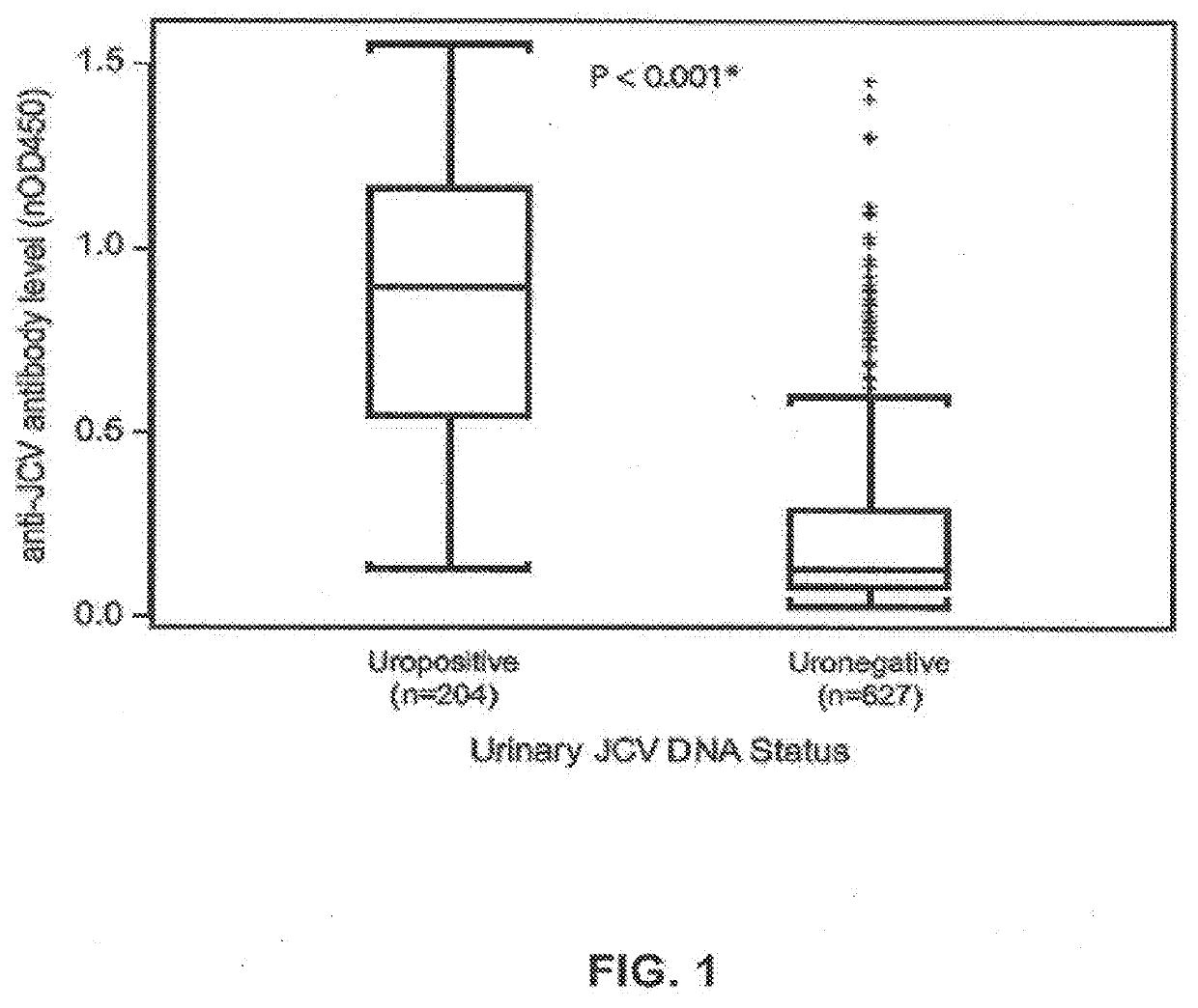
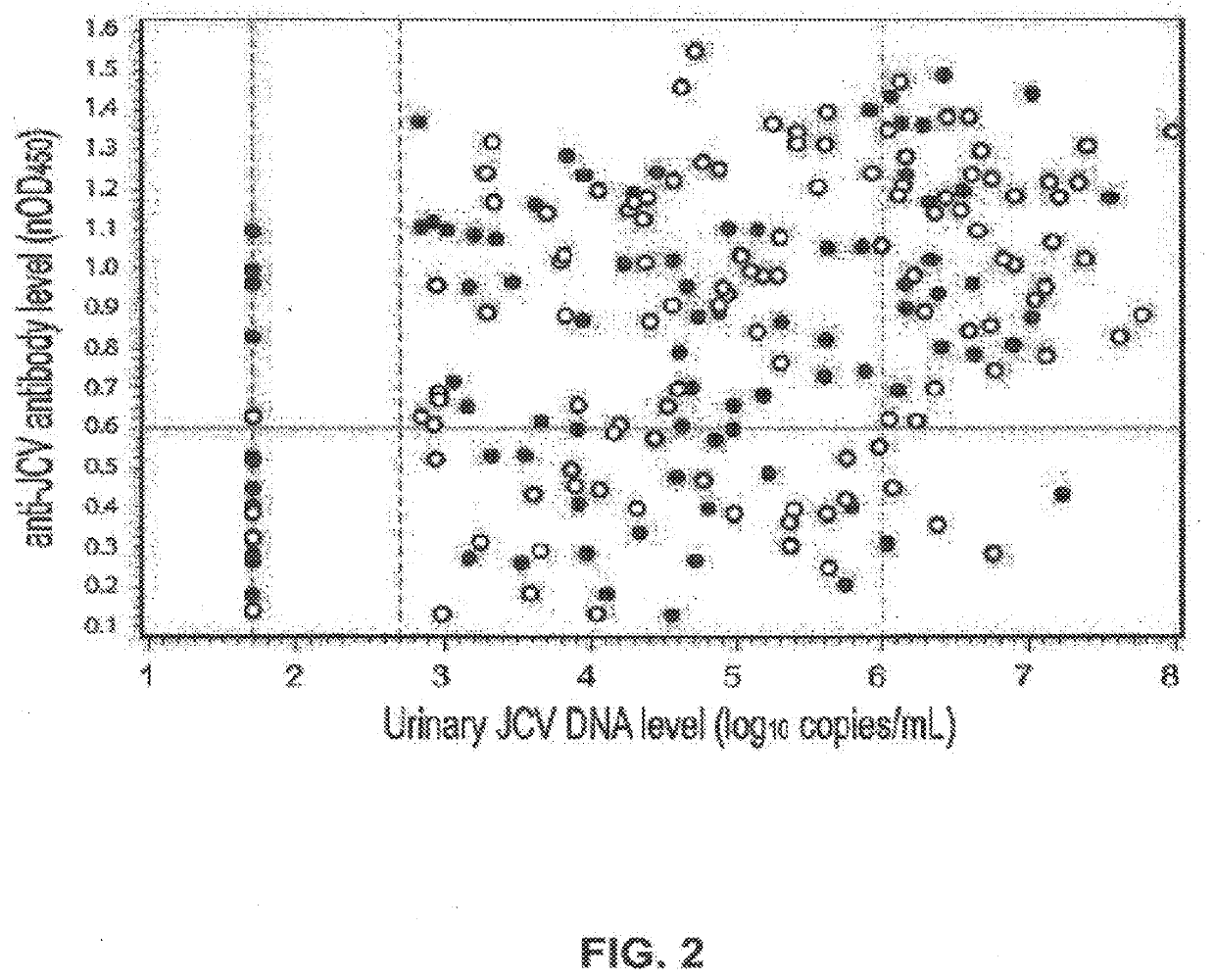
[0091]A sensitive assay for anti-JCV antibodies was developed using the HPVLPs described herein and is referred to herein in its various embodiments as an HPVLP assay. In an example of the assay, 96 well microtiter plates were prepared by adding a solution containing HPVLP at a concentration of 1 μg / ml and incubating the plate overnight at 4° C. The wells were rinsed with diluent buffer and then blocked for one hour at room temperature with Casein Blocking Buffer and rinsed with diluent buffer. The assay controls and serum or plasma samples were diluted 1:200 in assay diluent. The diluted samples and controls were added to wells and incubated for one hour at room temperature and washed with diluent buffer. Detection was performed using donkey anti-human-HRP antibody (IgG), which was added to the wells and incubated at room temperature for one hour. Plates were then washed and TMB (3,3′,5,5′-tetramethylbenzidine) buffer (Chromagen, Inc., San Diego, Calif.) was added. After...
example 3
Confirmation Assay
[0097]In some cases, a secondary confirmation assay (secondary assay) was carried out in addition to the test described supra. In the confirmation assay, samples (plasma or serum) were incubated with HPVLP (final VLP concentration=1 μg / mL; final sample dilution=1:200) for one hour at room temperature prior to use in the assay. Control samples were incubated in assay buffer, and not in the presence of HPVLP. The assay was then conducted as described above. A percent nOD450 inhibition was calculated as: % inhibition=100×[1−(average nOD450) (JCV MAD-1 VLP pre-incubated samples)÷(average nOD450) (buffer incubated samples)].
[0098]If the assay results were the same after pre-incubation with buffer as in the primary assay (i.e., approximately the same O.D.), then the sample was interpreted to be negative for the presence of JCV-specific antibodies. If the assay results were lower after pre-incubation with HPVLPs (i.e., in the secondary assay), then the sample was interpre...
PUM
 Abstract
Description
Claims
Application Information
 - R&D
- Intellectual Property
- Life Sciences
- Materials
- Tech Scout
- Unparalleled Data Quality
- Higher Quality Content
- 60% Fewer Hallucinations
Browse by: Latest US Patents, China's latest patents, Technical Efficacy Thesaurus, Application Domain, Technology Topic, Popular Technical Reports.
© 2025 PatSnap. All rights reserved.Legal|Privacy policy|Modern Slavery Act Transparency Statement|Sitemap|About US| Contact US: help@patsnap.com


