Method, system and computer readable medium for automatic segmentation of a 3D medical image

a medical image and automatic segmentation technology, applied in the field of image processing, can solve the problems of insufficient reliability, time-consuming and laborious, and manual segmentation of outlines on a contiguous set of 2d slices, and achieve the effect of reducing processing time and/or resources and efficient localization

Active Publication Date: 2021-04-29
GE PRECISION HEALTHCARE LLC
View PDF7 Cites 3 Cited by
  • Summary
  • Abstract
  • Description
  • Claims
  • Application Information

AI Technical Summary

Benefits of technology

[0020]An automatic segmentation method according to the subject matter disclosed herein can decrease the processing time and / or resources and make possible to establish an efficient localization of the bounding box of the region of interest. The automatic segmentation disclosed herein is proved to be accurate and robust not requiring any post editing, and successfully eliminating the problems related to inter- and intra-operator variability.

Problems solved by technology

Reluctance to use the fully automatic approach is due to the concerns about its insufficient reliability in cases where the target anatomy may difference from the norm, as well as due to high computational demands of the approach based on image registration.
Manually tracing the outlines on a contiguous set of 2D slices and then combining them can be time consuming and labor intensive.
Some organs are located in a soft tissue environment wherein resolution against surrounding structures has poor contrast since neighboring organs have similar density values.
As mentioned above, at this moment manual contouring is the gold standard for organ segmentation, which requires contouring various structures slice by slice, which is a time consuming, expensive activity and can be inaccurate due to human errors.
The need for precise segmentation tools is unquestionable.
Alzheimer's disease gradually leads to nerve cell death and tissue loss throughout the brain.
Over time, the brain shrinks dramatically, affecting nearly all its functions.
The difficulties in segmenting smaller organs inside the brain is the image resolution and the low contrast between neighboring tissues, so one can mainly trust on relative localization based on larger, better separable organs; for example, the ventricle is relevant as the examined organs of the basal ganglia are located next to it.
The drawback of this solution is that both of the two stages are carried out in 3D, resulting in a high computational demand; the coarse-scale segmentation, which is only a preliminary or preparational stage of the effective segmentation stage necessitates considerable computer resources due to its 3D character.
These known methods do not solve the problems of resource-efficient and reliable automatic segmentation, especially for segmentation of brain MR images, where the above mentioned difficulties arise.

Method used


Image

  • Method, system and computer readable medium for automatic segmentation of a 3D medical image
  • Method, system and computer readable medium for automatic segmentation of a 3D medical image
  • Method, system and computer readable medium for automatic segmentation of a 3D medical image

Examples


Embodiment Construction

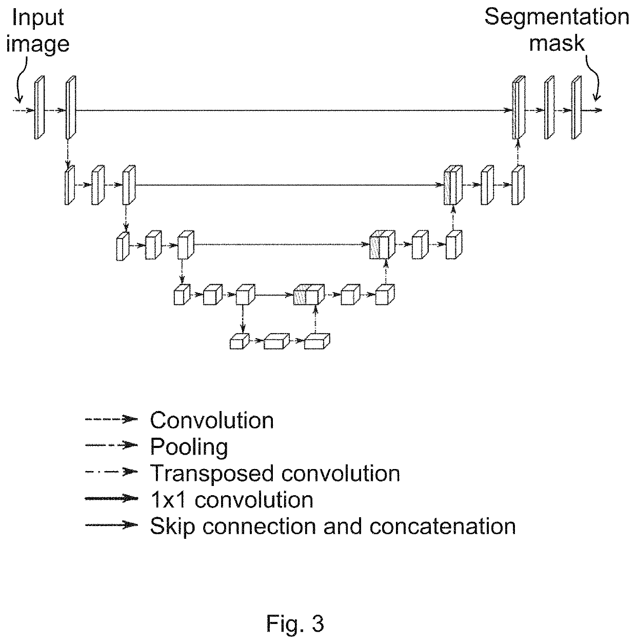
[0027]The subject matter disclosed herein is a method based on a machine learning model, e.g. on a deep-learning technique. In the following, an exemplary image dataset is described for use in the method, as well as an exemplary suitable architecture, and exemplary details of the training.

Image Dataset

[0028]The exemplary image dataset was collected for a study on Alzheimer's disease. All images were standard T1 images (T1-weighted scans produced by MRI using short Time to Echo and Repetition Time values) depicting the brain and small part of the neck. All images were preprocessed with an available tool that transforms images into a common geometry (orientation, resolution, uniform voxel size) and applies atlas to segment several structures in the brain. The automatically segmented contours (used for both model training and evaluation) were not verified by medical doctors, so they can be only considered ‘pseudo-gold’ segmentations.

[0029]Additional pre-processing was applied—which is ...


PUM


Abstract

A method, a system and a computer readable medium for automatic segmentation of a 3D medical image, the 3D medical image comprising an object to be segmented, the method characterized by comprising: carrying out, by using a machine learning model, in at least two of a first, a second and a third orthogonal orientation, 2D segmentations for the object in slices of the 3D medical image to derive 2D segmentation data; determining a location of a bounding box (10) within the 3D medical image based on the 2D segmentation data, the bounding box (10) having predetermined dimensions; and carrying out a 3D segmentation for the object in the part of the 3D medical image corresponding to the bounding box (10).

Description

FIELD OF THE INVENTION[0001]The subject matter disclosed herein relates to a method, a system and a computer readable medium for automatic segmentation of a 3D medical image using a machine learning model. More particularly, the disclosed subject matter relates to image processing, and to systems and methods for medical imaging with efficient and accurate three-dimensional 3D image segmentation capabilities.BACKGROUND OF THE INVENTION[0002]Segmentation of anatomical structures, e.g. organs in 3D medical images is a fundamental task in a number of clinical processes in the field of oncology, radiology and in planning surgical interventions. A 3D medical image is a medical image of a 3D volume in a subject along an orientation such as axial, coronal, sagittal, or oblique, where the medical images may be acquired by a 2D acquisition, a 3D acquisition, or a combination thereof. These 3D medical images can be considered as a set of 2D slices or layers in each of the 3D directions, i.e. a...

Claims


Application Information

Patent Timeline
Patent Type & Authority Applications(United States)
IPC IPC(8): G16H30/40G06T7/00G06T7/10G06N3/08
CPCG16H30/40G06T7/0012G06T7/10G06T2210/12G06T2207/30004G06T2207/20081G06T2207/20084G06N3/08G06T7/11G06N3/045G06T2207/10088G16H50/20
Inventor RUSKO, LASZLOGRECCHI, ELISABETTATAKACS, PETRA
Owner GE PRECISION HEALTHCARE LLC
Why Eureka
Try Eureka