Eureka AIR delivers breakthrough ideas for toughest innovation challenges, trusted by R&D personnel around the world.

Endoscopic Photoacoustic Probe

Pending Publication Date: 2021-07-15
UNIV GRENOBLE ALPES
View PDF0 Cites 0 Cited by
  • Summary
  • Abstract
  • Description
  • Claims
  • Application Information

AI Technical Summary

Benefits of technology

The invention is a photoacoustic probe designed for arthroscopy. It comprises an inclined catheter with an optical fibre for laser coupling, an ultrasound sensor, and a control handle for easy handling. The probe has a sheath made of a special material allowing ultrasounds and laser to pass through. The first portion of the sheath has a constant thickness, while the second portion has a larger diameter. The invention also includes an imaging system connected to a laser source and an endoscopic photoacoustic probe for displaying the vascularization of an intra-articular area. The processor can also display an ultrasound image of the meniscus and a photoacoustic image of the vascularization of the meniscus.

Problems solved by technology

However, they represent an imperfect imaging panoply: some information remains inaccessible or not very user-friendly (notably not being in real time), and irradiations are recognised as a major problem for patients and medical staff.
During an arthroscopy, the visual exploration of the inside of the articular cavity is limited to the surface of the structures constituting the joint, with, as consequences, potential diagnostic or therapeutic limitations.
Furthermore, since it involves an inaccessible zone, it does not lend itself to the injection of a contrast agent or a fluorescent agent to show up the vascularisation of the anatomical structures.
However, the configuration of a joint and the space available for the insertion of such a probe make the use of photoacoustics in arthroscopy difficult.

Method used


Image

  • Endoscopic Photoacoustic Probe
  • Endoscopic Photoacoustic Probe
  • Endoscopic Photoacoustic Probe

Examples


Embodiment Construction

[0010]An aim of the invention is to conceive an endoscopic photoacoustic probe suited for arthroscopy.

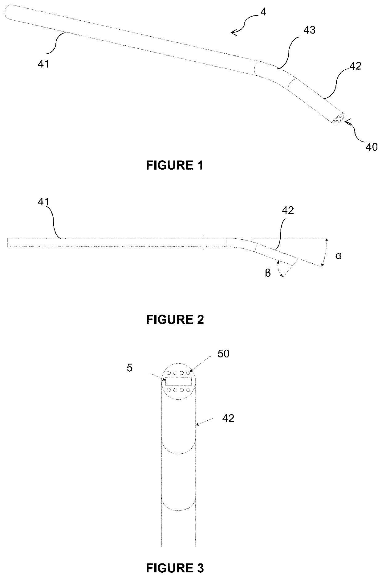
[0011]To this end, the invention proposes a photoacoustic ultrasound probe, comprising:[0012]a catheter,[0013]an ultrasound sensor arranged at a distal end of the catheter,[0014]at least one optical fibre suited to being connected to a laser source, said optical fibre extending into the catheter up to the distal end,

[0015]said probe being characterised in that the catheter has an inclined distal portion and / or a bevelled distal end.

[0016]In the present text, the relative terms “proximal” and “distal” are understood respectively to be a part of the probe situated on the side of the operator who handles it, and of a part of the probe on the side of the body of the patient into which it is intended to be inserted.

[0017]According to an embodiment, the catheter has a distal portion inclined by an angle comprised between 10 and 30° with respect to a proximal portion of the catheter. Accor...


PUM


Abstract

The invention relates to a photoacoustic ultrasound probe (100), comprising:a catheter (4),an ultrasound sensor (5) arranged at a distal end (40) of the catheter,at least one optical fibre (50) suited to being connected to a laser source, said optical fibre extending into the catheter up to the distal end (40),said probe being characterised in that the catheter has an inclined distal portion (42) and / or a bevelled distal end (40).

Description

FIELD OF THE INVENTION[0001]The present invention relates to an endoscopic photoacoustic probe.PRIOR ART[0002]Medical imaging groups together means for acquiring and rendering images from different physical phenomena: magnetic resonance, reflection of ultrasonic waves, radioactivity, absorption of X-rays, etc. These technologies make it possible to view the physiology or the metabolism of the human body, to establish a precise diagnosis and to orientate therapeutic choices. However, they represent an imperfect imaging panoply: some information remains inaccessible or not very user-friendly (notably not being in real time), and irradiations are recognised as a major problem for patients and medical staff.[0003]Only certain technologies are available in the operating theatre depending on the type of surgery. Orthopaedics requires the identification of bone structures and thus mainly resorts to the use of X-rays.[0004]The use of intraoperative imaging has the aim of providing additiona...

Claims


Application Information

Patent Timeline
IPC IPC(8): A61B5/00
CPCA61B5/0095A61B5/6852A61B2562/227A61B5/4528A61B5/4514A61B2505/05A61B8/12
Inventor CUSTILLON, GUILLAUMEDUBOIS, NICOLAS
Owner UNIV GRENOBLE ALPES
Why Eureka
Eureka Blog
Learn More