Method for detecting synovial joint infections

Pending Publication Date: 2021-08-26
QUALIZYME DIAGNOSTICS GMBH & CO KG
View PDF0 Cites 0 Cited by
  • Summary
  • Abstract
  • Description
  • Claims
  • Application Information

AI Technical Summary

Benefits of technology

[0011]It turned surprisingly out that a synovial joint infection can be detected by determining lysozyme activity in the synovial fluid of a mammal suffering from a synovial joint condition, wherein the enzymatic activity is preferably determined in an untreated synovial fluid sample obtained directly from said mammal (i.e. obtained by puncturing a synovial joint of a mammal). Until now it was thought that all kind of synovial joint conditions—either caused by infections or by non-infectious inflammation—result in an increase of lysozyme activity. However, as herein shown lysozyme activity in synovial fluid from mammals suffering from a non-infectious inflammatory condition of a synovial joint do not or not significantly show lysozyme activity. This finding is even more surprising considering that the activity of myeloperoxidase—a well known marke

Problems solved by technology

However, as herein shown lysozyme activity in synovial fluid from mammals suffering from a non-infec

Method used


Image

  • Method for detecting synovial joint infections
  • Method for detecting synovial joint infections
  • Method for detecting synovial joint infections

Examples


example 1

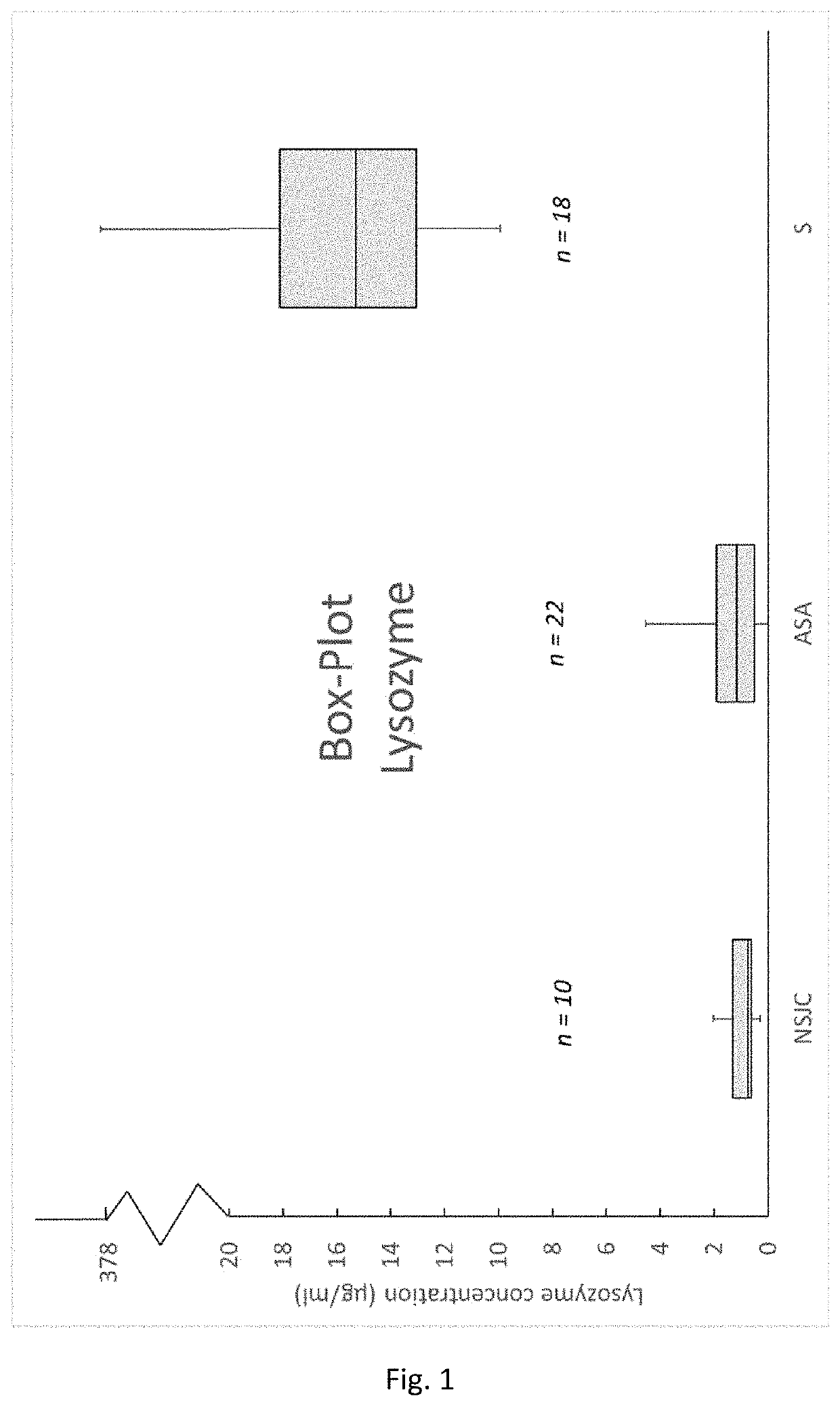
tion of Lysozyme Activity in Synovial Fluid Samples Obtained From Healthy Patients and Patients Suffering From Rheumatoid Arthritis and Microbial Infections of a Synovial Joint

[0075]Lysozyme is an enzyme, which is produced and secreted by cells of the innate immune system. Peptidoglycan is the main building block of cell walls from gram positive bacteria and can be cleaved by lysozyme. This is a natural defence mechanism from the immune system to cope with bacteria by digesting their cell walls. It was demonstrated, that in 100% (see Table 1, Sensitivity) of the tested synovia samples which were diagnosed as septic by the responsible clinicians, the lysozyme activity was elevated. Moreover, 100% (see Table 1, Specificity) of the samples which were classified as negative or inflammatory did not show elevate lysozyme levels.

[0076]The activities of lysozyme were measured as follows:

[0077]290 μl of a 0.45 mg / ml peptidoglycan solution (from Micrococcus luteus) in a sodium phosphate buffe...

example 2

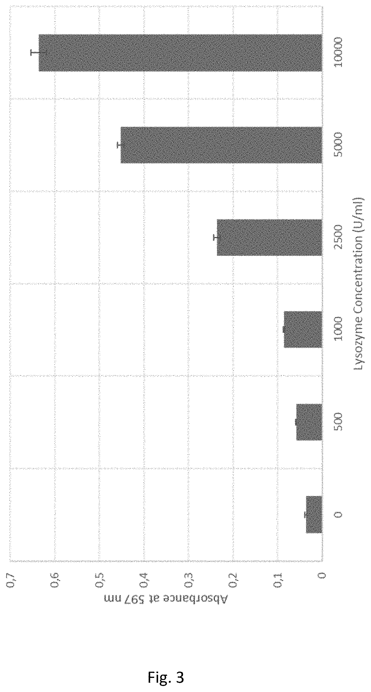
Measurement of Lysozyme With Dyed Peptidoglycan

[0078]7.5 mg peptidoglycan-reactive black 5 were suspended in 0.990 ml 0.9% NaCl solution. 10 μl of different lysozyme concentrations were added to the reaction solution to final concentrations ranging from 0 U / ml-10000 U / ml (every concentration in triplicates). All samples were incubated at 37° C. for 60 minutes and shaking at 1400 rpm. After the incubation step all samples were centrifuged at 10000 g for 5 minutes. 100 μl of each supernatant were transferred into a 96 well plate and absorbances were measured at 597 nm. Absorbance values are shown in FIG. 3. All lysozyme concentrations could be clearly distinguished.

example 3

se Over 60 Minutes for 10000 U / ml Lysozyme

[0079]7.5 mg / ml peptidoglycan-reactive black 5 were suspended in 0.990 ml 0.9% NaCl solution. 10 μl of a lysozyme solution were added to a final concentration of 10000 U / ml. All samples were incubated at 37° C. for 10-60 minutes and 1400 rpm. Every 10 minutes triplicates were centrifuged (10000 g, 5 min) and 100 μl of the supernatants were transferred into a 96 well plate. The absorbance was measured at 597 nm. The time-depending dye release is shown in FIG. 4. Already after 10 minutes the


PUM


Abstract

The present invention relates to a method for detecting a synovial joint infection comprising the steps of
    • contacting a synovial fluid sample with at least one lysozyme substrate, and
    • detecting a synovial joint infection when a conversion of said at least one substrate is determined

Description

TECHNICAL FIELD[0001]The present invention relates to methods for detecting infections in synovial joints.BACKGROUND ART[0002]The infection of joints such as knees, hips or shoulders is usually the result of a surgical procedure or injuries in which bacteria or viruses can enter the joints. Thus, artificial joints or implants are often affected by such infections. The detection of a bacterial infection, for instance, in joints is carried out by means of a joint puncture with synovial analysis, whereby the bacteria are detected by culturing on special culture media in an incubator. However, a negative culture result does not prove that it is free of any germs, as there are up to 30% false negative tests (Simank HG et al. (2004) Joint Anemia Orthopedist 33: 327-31). This microbial cultivation detection system usually takes 2 to 5 days. Alternatively, a leukocyte count can be performed in the synovial fluid (leukocyte / mm3). However, leukocyte counts between 1,000 and 10,000 do not prec...

Claims


Application Information

Patent Timeline
IPC IPC(8): C12Q1/34C12Q1/28
CPCC12Q1/34G01N2800/10G01N2333/936C12Q1/28
Inventor HEINZLE, ANDREAGAMERITH, CLEMENSLUSCHNIG, DANIELSIGL, EVA
Owner QUALIZYME DIAGNOSTICS GMBH & CO KG
Why Eureka
Try Eureka