Eureka AIR delivers breakthrough ideas for toughest innovation challenges, trusted by R&D personnel around the world.

Method and apparatus for separation of tissue

a tissue and method technology, applied in the field of tissue separation methods and equipment, can solve the problems of no assistive, automated, guided system for tissue preparation, etc., and achieve the effect of facilitating better clinical outcomes in dalk, easy and clean tissue separation, and facilitating the separation of layers

Pending Publication Date: 2022-01-06
EYEDEA MEDICAL INC
View PDF0 Cites 0 Cited by
  • Summary
  • Abstract
  • Description
  • Claims
  • Application Information

AI Technical Summary

Benefits of technology

The disclosed invention is an apparatus that helps to separate corneal tissue layers for use in surgery. It includes a stabilizing corneal base, pressure feedback system, and fluid injection system. The apparatus optimizes the positioning of a needle inserted into the cornea to create a fluid bubble that separates the Descemet's Membrane from the stroma, making the separation process easier and cleaner. The device also reduces training time, processing time, and increases accuracy and yield of the surgery. It helps to expand the donor pool while maintaining quality, making it easier to prepare corneal tissue grafts. This assistive, standardizing system allows technicians and surgeons to more easily and quickly separate corneal tissue layers, even in low-resource settings.

Problems solved by technology

There are no assistive, automated, or guided systems for preparing tissue.

Method used


Image

  • Method and apparatus for separation of tissue
  • Method and apparatus for separation of tissue
  • Method and apparatus for separation of tissue

Examples


Embodiment Construction

[0035]The present disclosed invention has many possible embodiments. The specific embodiments shown herein should not be construed as the final embodiments of the apparatus, but simply examples of different variations of the apparatus. The present invention has a large scope in that this apparatus can come in many forms. Similarly, the methods are examples of potential ways that the apparatus can be handled and used to perform tissue separation. However, the method of using an apparatus to prepare certain ocular tissues is novel and has some variations.

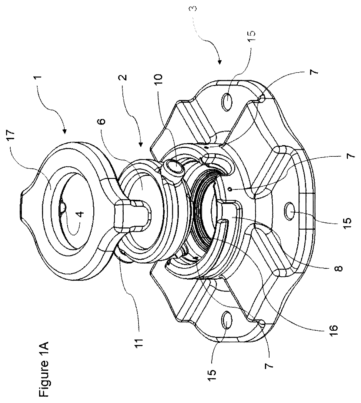
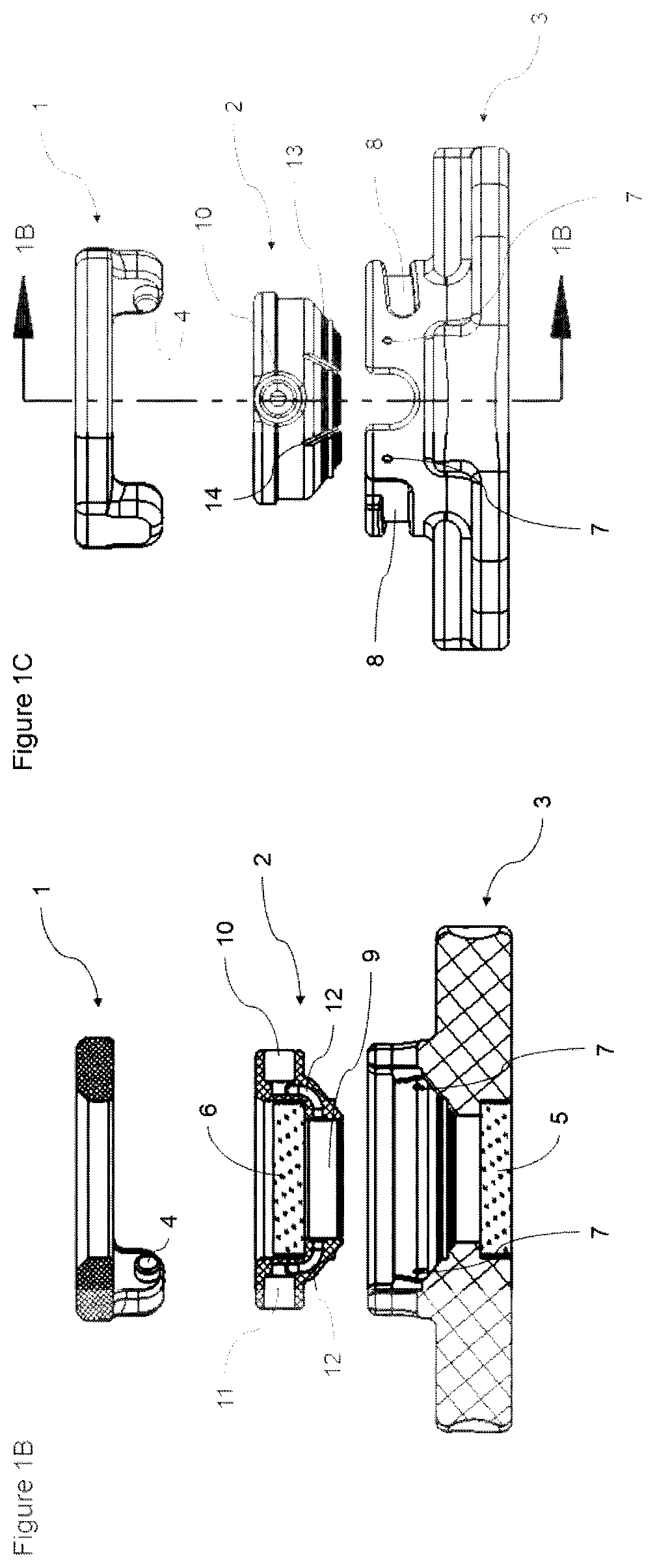
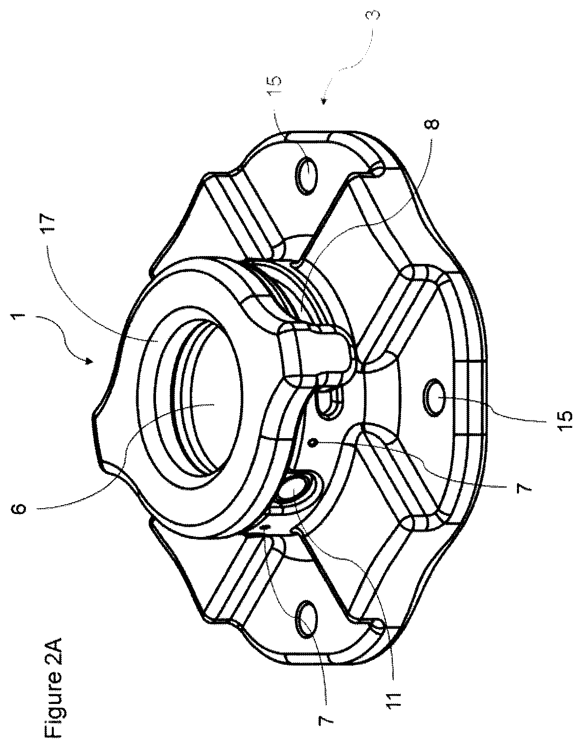
[0036]The novel, ocular preparation system or apparatus of the disclosed invention includes the base 27, the needle 28, the sensor feedback system 25, and controlled fluid injection 24. The base 27 of the system is meant to fit ocular tissue, including a cornea, and needs to fit under a microscope. The base of the system comprises a base part 3, a stabilizing part 2, and can include a locking part 1. These parts assembled result in th...


PUM


Abstract

An ophthalmic medical device for separating layers of eye tissue is disclosed. The device may include a supporting structure for supporting the eye and the supporting structure may have at least one stabilizing feature that stabilizes the eye to reduce movement. The supporting structure may define at least one hollow channel for positioning a needle that is inserted into the cornea to make a fluid bubble that separates the Descemet's Membrane away from the underlying stroma. In some embodiments, the fluid bubble enables an easy, clean separation of the layers to later be transplanted in patients needing corneal transplants.

Description

TECHNICAL FIELD[0001]The presently disclosed subject matter relates generally to the design of medical devices. More particularly, the present invention relates to the use of a fluid bubble to cleave tissue within the cornea or other tissue in the eye.BACKGROUND[0002]The cornea consists of five main layers (anterior to posterior): the epithelium, Bowman's layer, the stroma, Descemet's Membrane, and the endothelium. The epithelium is the outermost layer of the surface of the eye, usually contains about five to seven layers of cells (50 microns thick), regenerates quickly, and acts as a barrier to the outside world. The Bowman's layer is a transition layer between the epithelium and stroma; this layer is usually only 8 to 14 microns thick and is largely involved in maintaining the proper shape of the cornea. The stroma is the bulk of the cornea, measuring around 500 microns thick and comprising of 90% of the cornea. This layer is filled with collagen fibers that are extremely regular ...

Claims


Application Information

Patent Timeline
IPC IPC(8): A61F9/007A61B90/11A61B90/14
CPCA61F9/007A61B2017/32035A61B90/14A61B90/11A61B2017/3405A61B17/3403A61B2017/3407
Inventor SOLLEY, KATHERINEOSTRANDER, BENJAMINDIAZ, CHRISTINEHAILEYESUS, AILONDOU, YUEHENGRUPPEL, III, EDWARDPARIKH, KUNAL
Owner EYEDEA MEDICAL INC
Why Eureka
Eureka Blog
Learn More