


Quick Research
Generate reliable direction feasibility study reports for your R&D in just a few steps.

Technical Q&A
Discover and master advanced knowledge NOW. Basics, ideas, possibilities, all at once.

Find Solutions
As an expert in R&D theories, this can generate solutions to your technical problems instantly.

Evaluate Feasibility
Analyze your overall solution with one click, know your potential R&D risks in advance.

Monitor Landscape
Get weekly tech updates, stay abreast of the latest tech innovations and key insights.
Marker for diagnosing neurodegenerative disease, and therapeutic composition
a neurodegenerative disease and marker technology, applied in the field of neurodegenerative disease markers, can solve the problems of lack of research on a marker or composition related to fus protein diagnosis, lack of knowledge about the mechanisms involved, fatal muscle atrophy and paralysis, etc., to achieve the effect of effectively preventing, treating or alleviating a neurodegenerative disease, and inhibiting brain cytoplasm aggregation and neurocytotoxicity
- Summary
- Abstract
- Description
- Claims
- Application Information
AI Technical Summary
Benefits of technology
Problems solved by technology
Method used
Image


Examples
example 1
t Preparation and Experimental Methods
[0116]1-1. Preparation of Drosophila melanogaster
[0117]All Drosophila stocks were stored under a standard food condition, a normal temperature (25° C.) and a normal humidity condition (60%), and Drosophila crossing was carried out according to standard procedures, and all offspring were raised at 25° C.
[0118]1-2. Cell Culture
[0119]Mouse neuroblastoma, Neuro2a cells, were cultured in DMEM (Life Technologies) containing 10% fetal bovine serum (FBS; Gibco) and a penicillin-streptomycin solution (50 mg / mL; Gibco) at 37° C. and 5% CO2 / 95% air conditions.
[0120]1-3. In Vitro Glutathionylation Assay
[0121]The full-length protein of myc-tag human FUS (0.51 μg) purified from HEK293 (OriGene) cells was incubated in 50 mM Tris-HCl (pH 7.5) in the presence of various concentrations of an oxidized glutathione (glutathione disulfide, GSSG) at 25° C., and one hour after incubation, the sample was placed on ice, and 3× non-reducing LDS sample buffer (Invitrogen)...
example 2
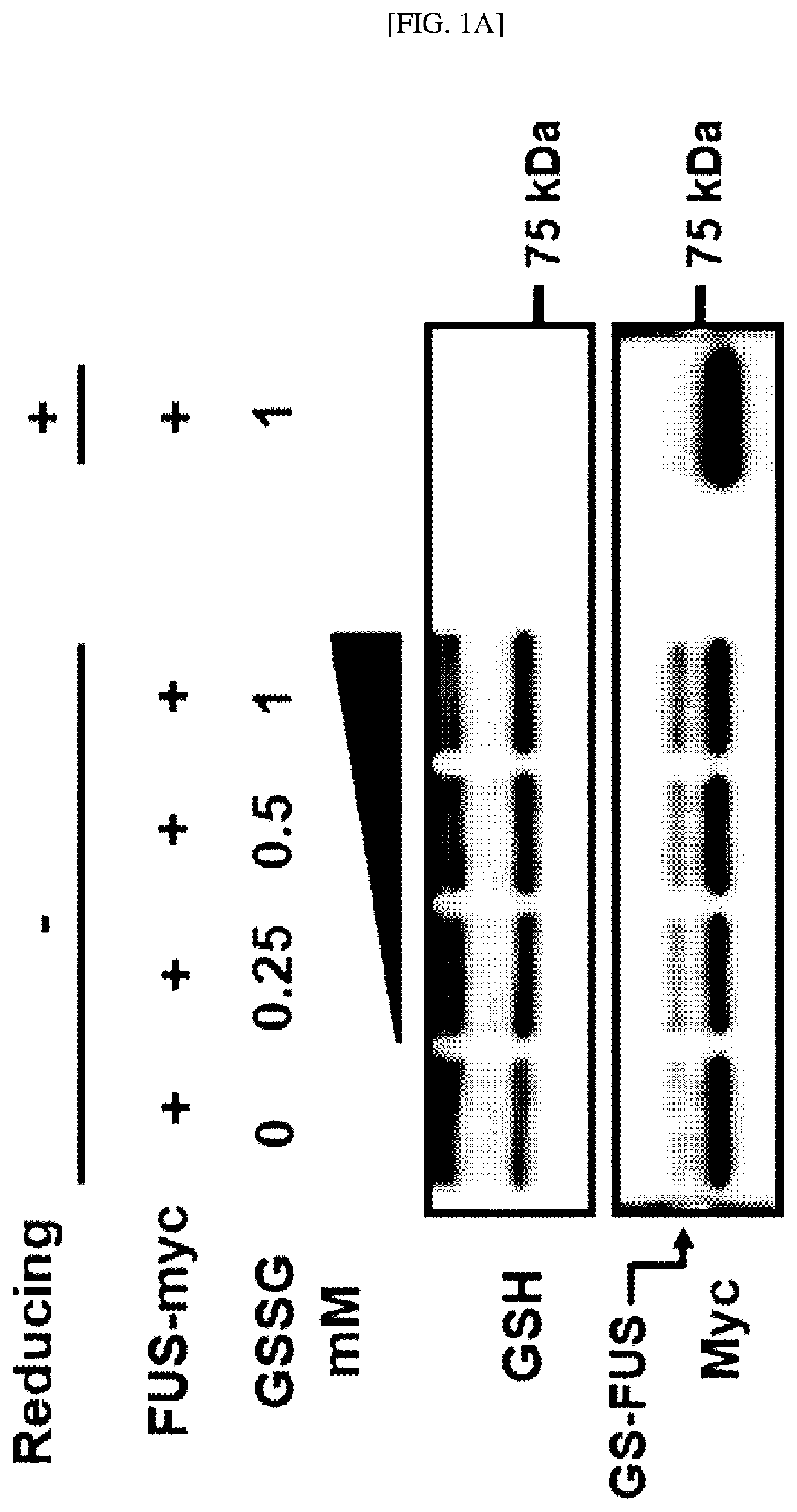
ion of In Vitro Glutathionylation of FUS Protein
[0172]In this example, to confirm whether in vitro glutathionylation of a FUS protein occurs, the oxidized glutathione (glutathione disulfide, GSSH) was added to a myc-labeled human FUS recombinant full-length protein and incubated, separation through sodium dodecyl sulfate-polyacrylamide gel electrophoresis (SDS-PAGE) was performed, and western blotting analysis was performed using a mouse anti-GSH antibody and a mouse anti-myc antibody.
[0173]As a result, as shown in FIGS. 1A and 1B, as the concentration of the oxidized glutathione (Glutathione disulfide, GSSH) increases (0 mM, 0.25 mM, 0.05 mM and 1 mM), it was confirmed that a larger amount of a glutathionylated FUS protein is detected. From the result, it was confirmed that the glutathionylation of a FUS protein occurs in vitro depending on the concentration of the oxidized glutathione (glutathione disulfide, GSSH).
example 3
ion of In Vivo Glutathionylation of FUS Protein
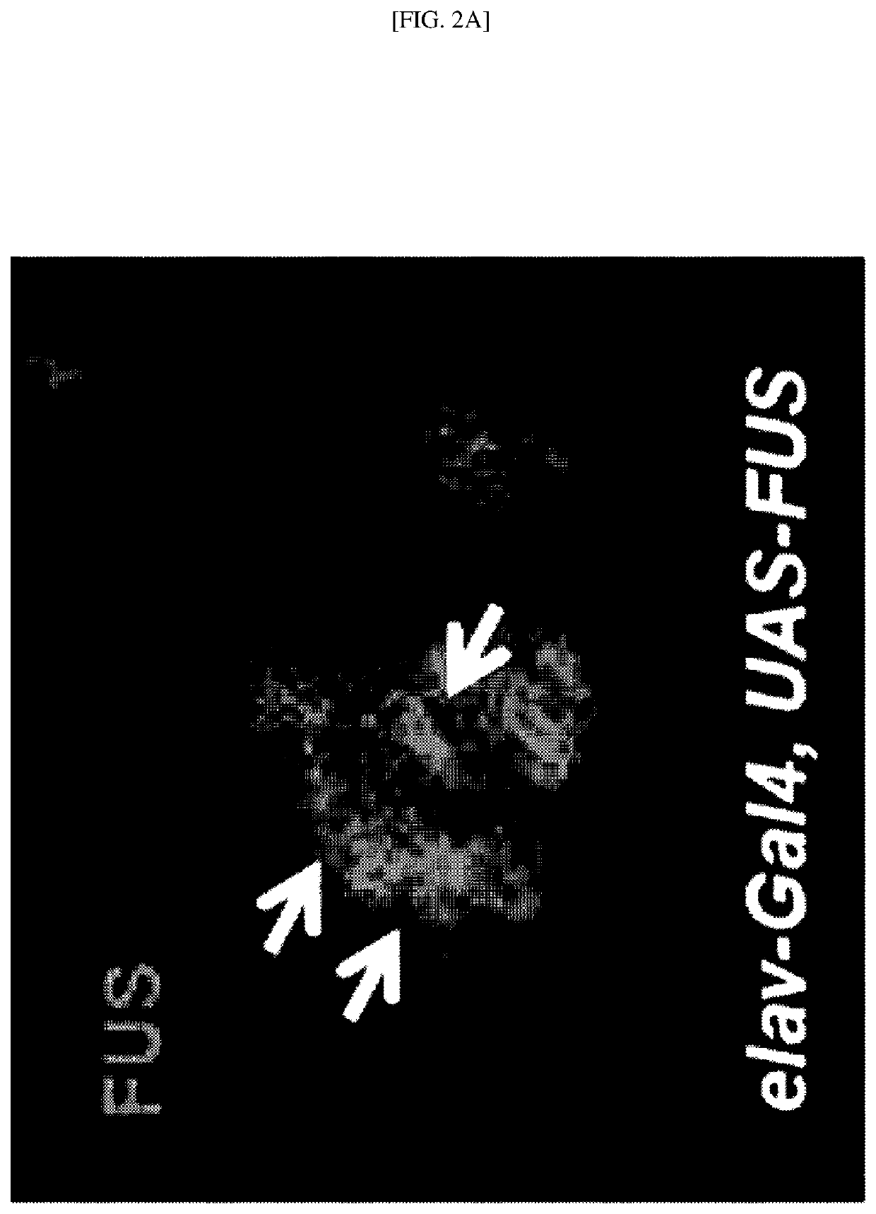
[0174]To confirm whether the glutathionylation of FUS protein occurs in vivo, a human FUS protein specifically expressed in neurons was expressed in Drosophila neurons using an elav-Gal4 driver. Specifically, a pCMV6-FUS-GFP-labeled human wild-type FUS protein was transfected into Drosophila. A region stained with DAPI represents the location of the nucleus.
[0175]As a result, the human wild-type FUS protein is located mainly in the nucleus of neurons of the brain of Drosophila, but as indicated by the arrows of FIG. 2A, when the human wild-type FUS protein is overexpressed, it was confirmed that a large amount of human wild-type FUS protein aggregates are observed in the cytoplasm (green). In addition, as indicated by the arrows of FIG. 2B, it was confirmed that the reduced glutathione (GSH) is observed in the cytoplasm (red). In addition, as indicated by the arrows of FIG. 2C in which FIGS. 2A and 2B are combined, it was confirmed that...
PUM
| Property | Measurement | Unit |
|---|---|---|
| Composition | aaaaa | aaaaa |
| Level | aaaaa | aaaaa |
Abstract
Description
Claims
Application Information
 - R&D Engineer
- R&D Manager
- IP Professional
- Industry Leading Data Capabilities
- Powerful AI technology
- Patent DNA Extraction
Browse by: Latest US Patents, China's latest patents, Technical Efficacy Thesaurus, Application Domain, Technology Topic, Popular Technical Reports.
© 2024 PatSnap. All rights reserved.Legal|Privacy policy|Modern Slavery Act Transparency Statement|Sitemap|About US| Contact US: help@patsnap.com