Enhancing epithelial or endothelial barrier function
a technology of epithelial or endothelial barrier and function, which is applied in the direction of transferases, peptide/protein ingredients, drug compositions, etc., can solve the problems of poorly understood epithelial injury and repair mechanisms
- Summary
- Abstract
- Description
- Claims
- Application Information
AI Technical Summary
Benefits of technology
Problems solved by technology
Method used
Image
Examples
example 1
Material and Methods
[0129]This example descibes material and methods used in Examples 2-9 bellow.
Plasmids and siRNA
[0130]The plasmids used in this study were obtained from a range of sources. pCDNA3 and pCMV-EGFP plasmids were purchased from INVITROGEN (Carlsbad, Calif.). Mouse Na+, K+-ATPase β2 subunit and mouse Na+, K+-ATPase β3 subunit with Myc-DDK tag were obtained from ORIGENE (Rockville, Md.). The Tet-On 3G drug-inducible gene expression system was purchased from CLONTECH (Mountain View, Calif.). The human Na+, K+-ATPase β1 subunit-coding sequence was inserted into the pTRE3 G vector at the Sall and BamHI restriction enzyme sites. The human MRCKα plasmid was a gift from Dr. Paolo Armando Gagliardi from the University of Bern (31). Knockdown was carried out using the TRIFECTA DSIRNA Kit (IDT, Coralville, Iowa) according to manufacturer's instruction. 100 nM of siRNA was used for each one million cells.
Antibodies and Inhibitors
[0131]The primary antibodies for western blot includ...
example 2 β1
Subunit Overexpression Increases Expression of Alveolar Tight Junctions
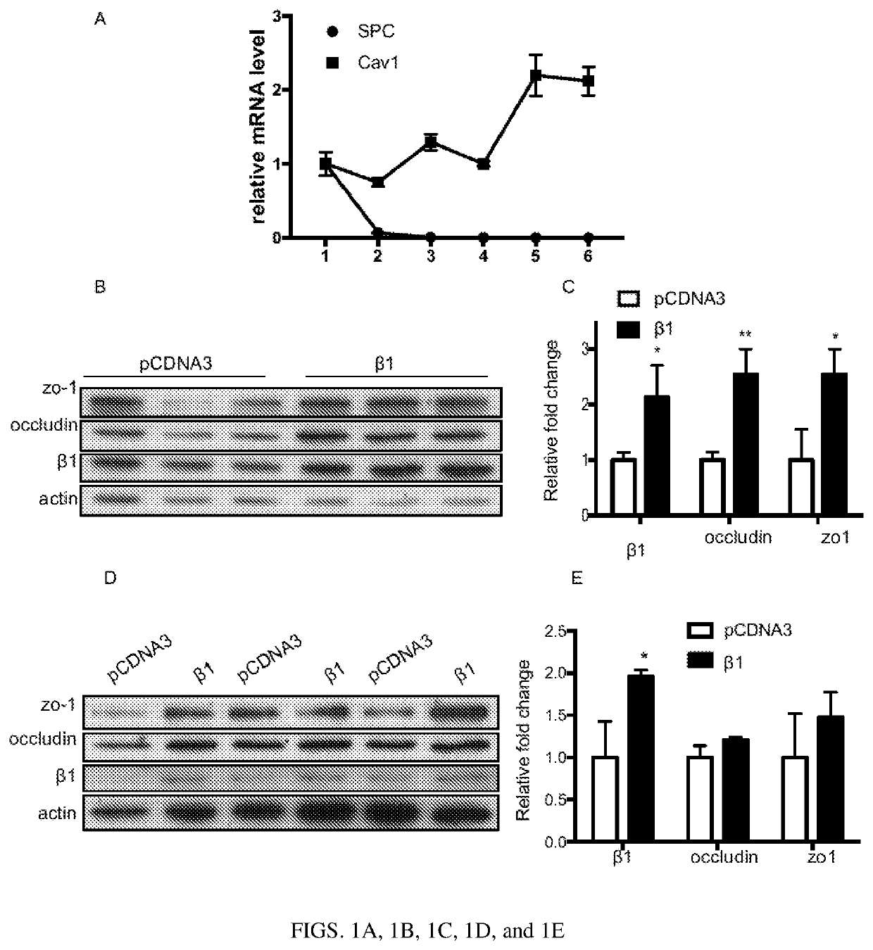
[0141]The majority of cell junctions in alveolar epithelial barrier are between adjacent ATI cells, which cover 95% of the its surface (5). Unfortunately, existing cell lines do not fully recapitulating the genetic and phenotypic characteristics of ATI in vivo and isolating ATI cells directly poses technical challenges (19). To overcome this, rat primary ATII cells were used since they are capable of differentiating into ATI cells when isolated and cultured in vitro (20). To track the phenotypic changes during the process, qPCR analysis was carried out for genes that are specific for ATII (SPC) or ATI (T1a) (FIG. 1A). SPC mRNA levels dropped 15-fold from 24 hours to 48 hours after isolation, suggesting a loss of ATII phenotype. In contrast, T1α level increased continuously until day five after isolation, indicating a shift to ATI phenotype. When cultured in transwell plates coated with 20 μg / ml fibronectin, these...
example 3
Overexpression of β1 Subunit Increases Alveolar Type I Barrier Integrity
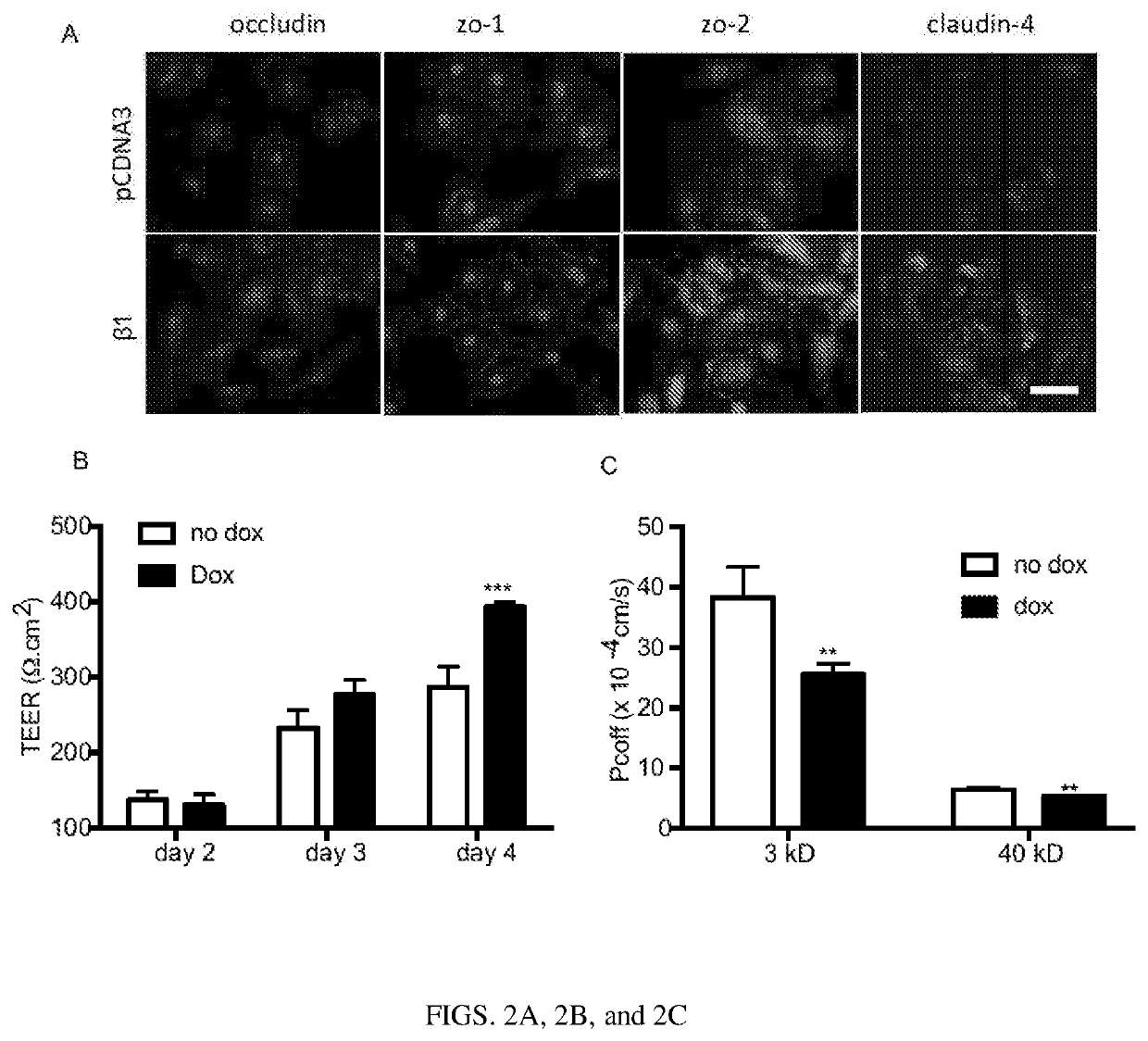
[0143]Given that the β1 subunit increased protein expressions of tight junctions in ATI cells, their localization in these cells was analyzed. Immunofluorescence staining confirmed increased level of occludin, zo-1, zo-2 and claudin-4 on the cell membrane (FIG. 2A). Next, assays were carried out to investigate the functional effect of the β1 subunit on the alveolar epithelial barrier. Since electroporation requires trypsinizing the cells, which would disrupt the cell monolayer and impede TEER and permeability assays, a doxycycline-inducible system was created to control the β1 subunit expression by cloning the rat β1 subunit into a Tet-on plasmid. This would allow one to turn on the gene expression in ATI cells at a later point after electroporation by just adding doxycline to the cell culture media. To test whether this system can be used to control the expression of the β1 subunit, 16HBE14o-cells, a human bron...
PUM
| Property | Measurement | Unit |
|---|---|---|
| temperature | aaaaa | aaaaa |
| surface area | aaaaa | aaaaa |
| pH | aaaaa | aaaaa |
Abstract
Description
Claims
Application Information
 - R&D
- Intellectual Property
- Life Sciences
- Materials
- Tech Scout
- Unparalleled Data Quality
- Higher Quality Content
- 60% Fewer Hallucinations
Browse by: Latest US Patents, China's latest patents, Technical Efficacy Thesaurus, Application Domain, Technology Topic, Popular Technical Reports.
© 2025 PatSnap. All rights reserved.Legal|Privacy policy|Modern Slavery Act Transparency Statement|Sitemap|About US| Contact US: help@patsnap.com


