Neural netork based identification of areas of interest in digital pathology images

a neural network and pathology image technology, applied in image enhancement, medical communication, instruments, etc., can solve the problems of lossless or lossy compression algorithm, difficult to detect morphological changes due to a test compound, tedious and error-prone process, etc., and achieve faster pathologist's review. , the effect of more reliabl

Pending Publication Date: 2022-03-10
LEICA BIOSYST IMAGING
View PDF0 Cites 7 Cited by
  • Summary
  • Abstract
  • Description
  • Claims
  • Application Information

AI Technical Summary

Benefits of technology

[0073]In summary of this aspect of the disclosure, a method, apparatus and system are provided for processing a histological image of a tissue sample of a given tissue type that has been treated with a test compound in order to detect toxic reactions to the test compound. An autoencoder is used that has been trained with a training data set comprising histological images of tissue samples which are of the given tissue type, but which have not been treated with the test compound. The trained au

Problems solved by technology

Since the range of normal tissue variation can be quite wide, this is not a straightforward task.
Despite the efficiency improvements introduced by digital pathology, detecting morphological changes due to a test compound remains a tedious and error-prone process

Method used


Image

  • Neural netork based identification of areas of interest in digital pathology images
  • Neural netork based identification of areas of interest in digital pathology images
  • Neural netork based identification of areas of interest in digital pathology images

Examples


example

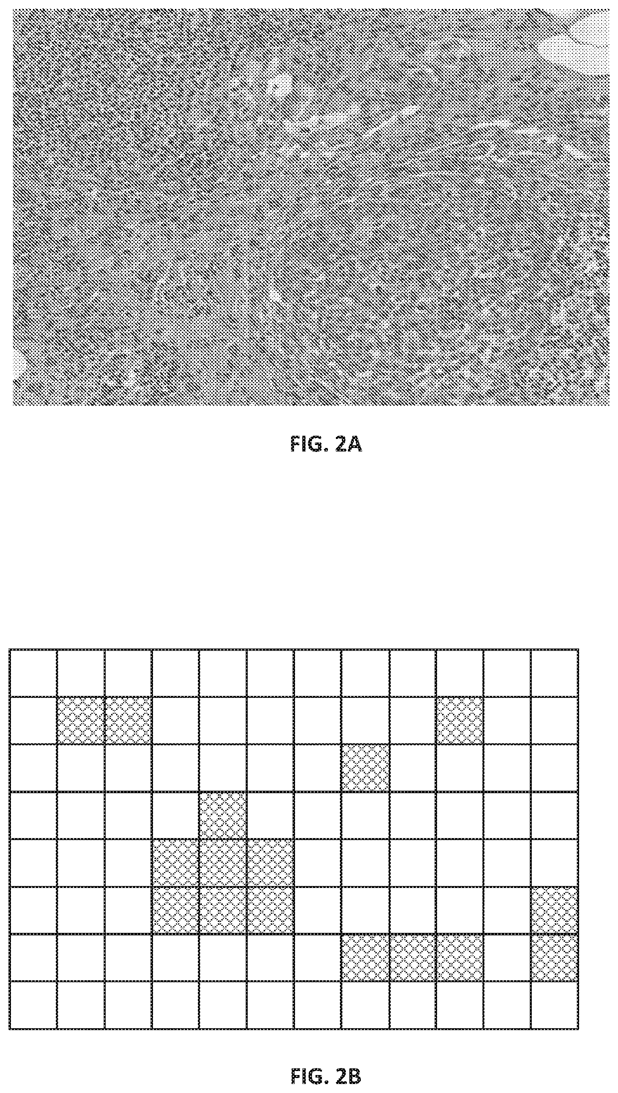
[0144]FIG. 2A shows a portion of a raw image as a patch from an H&E-stained WSI in which the cluster of larger, dark purple cells in the bottom right quadrant is a tumor, while the smaller dark purple cells are lymphocytes.

[0145]FIG. 2B shows the predicted areas of interest generated by the CNN as a mask. FIG. 2B is a schematic representation of a binary mask as could be generated by our CNN where the binary mask is based on non-overlapping image tiles, with the cross-hatched tiles being the areas of interest and the unhatched tiles are areas that are predicted not to be of interest. It will be understood that if a tile-based approach is taken, the number of tiles would typically much higher than the 12×8 array shown, e.g. by an order of magnitude. In other examples, the areas of interest may have their perimeters configured to follow a shape-based segmentation algorithm, and so represent an arbitrary shape rather than squares or rectangles.

[0146]Acquisition & Image Processing

[0147]...

example embodiment

[0269]In one embodiment, a method employed by computer-automated system for processing a histological image of a tissue sample that has been treated with a test compound begins with receiving the histological image of the tissue sample, the histological image including a two-dimensional array of pixels. Next, the system applies a first convolutional neural network to the histological image to generate a first output image patch with a two-dimensional array of pixels with a mapping to that of the histology image. The first output image patch is generated by assigning one of a plurality of relevance classes to each pixel, wherein the plurality of relevance classes includes at least one class representing a pixel of interest and at least one class representing a pixel that is not of interest. Advantageously, the first convolutional neural network has been trained using a training data set comprising histological images and pathologist interaction data, wherein the pathologist interacti...


PUM


Abstract

A CNN is applied to a histological image to identify areas of interest. The CNN classifies pixels according to relevance classes including one or more classes indicating levels of interest and at least one class indicating lack of interest. The CNN is trained on a training data set including data which has recorded how pathologists have interacted with visualizations of histological images. In the trained CNN, the interest-based pixel classification is used to generate a segmentation mask that defines areas of interest. The mask can be used to indicate where in an image clinically relevant features may be located. Further, it can be used to guide variable data compression of the histological image. Moreover, it can be used to control loading of image data in either a client-server model or within a memory cache policy. Furthermore, a histological image of a tissue sample of a tissue type that has been treated with a test compound is image processed in order to detect areas where toxic reactions to the test compound may have occurred. An autoencoder is trained with a training data set comprising histological images of tissue samples which are of the given tissue type, but which have not been treated with the test compound. The trained autoencoder is applied to detect tissue areas by their deviation from the normal variation seen in that tissue type as learnt by the training process, and so build up a toxicity map of the image. The toxicity map can then be used to direct a toxicological pathologist to examine the areas identified by the autoencoder as lying outside the normal range of heterogeneity for the tissue type. This makes the pathologists review quicker and more reliable. The toxicity map can also be overlayed with the segmentation mask indicating areas of interest. When an area of interest and an area identified as lying outside the normal range of heterogeneity for the tissue type, and increased confidence score is applied to the overlapping area.

Description

BACKGROUNDField of the Invention[0001]The present disclosure relates to image processing of pathology images with a neural network in order to find areas of clinical interest and areas including toxicological pathology.Related Art[0002]Digital pathology continues to change the way pathologists view and diagnose slides. The traditional way for pathologists to examine a slide is to observe a glass slide under a microscope. The pathologist will start by viewing the slide with a low magnification objective. When an area with potential diagnostic value is observed, the pathologist will switch to a high magnification objective to look in more detail at that area. Subsequently, the pathologist will switch back to low magnification to continue examining other areas on the slide. This low-high-low magnification viewing sequence may be repeated several times over the slide until a definite and complete diagnosis can be made for the slide.[0003]In the past twenty years, the introduction of dig...

Claims


Application Information

Patent Timeline
IPC IPC(8): G06T7/00G06K9/00G06K9/32G16H80/00G06V10/764
CPCG06T7/0012G06K9/0014G06T2207/10056G16H80/00G06T2207/20084G06K9/3233G06T7/11G06N3/08G06T2207/20081G06T2207/30096G06T2207/20021G06T2207/10024G06N3/045G06F18/2415G06V2201/03G06V10/26G06V10/945G06V10/82G06V20/695G06V10/454G06V10/764G06V2201/10
Inventor GEORGESCU, WALTERSALIGRAMA, KIRANOLSON, ALLENMALLYA UDUPI, GIRISHOLIVEIRA, BRUNO
Owner LEICA BIOSYST IMAGING
Why Eureka
Try Eureka