

Systems and Methods for Digital Pathology
a digital pathology and pathology technology, applied in the field of image processing, can solve the problems of time-consuming and laborious process of visual inspection of a roi on a microscope slide to identify potentially relevant pathology features, and achieve the effect of improving ease and speed
- Summary
- Abstract
- Description
- Claims
- Application Information
AI Technical Summary
Benefits of technology
Problems solved by technology
Method used
Image

Examples
Embodiment Construction
System Architecture
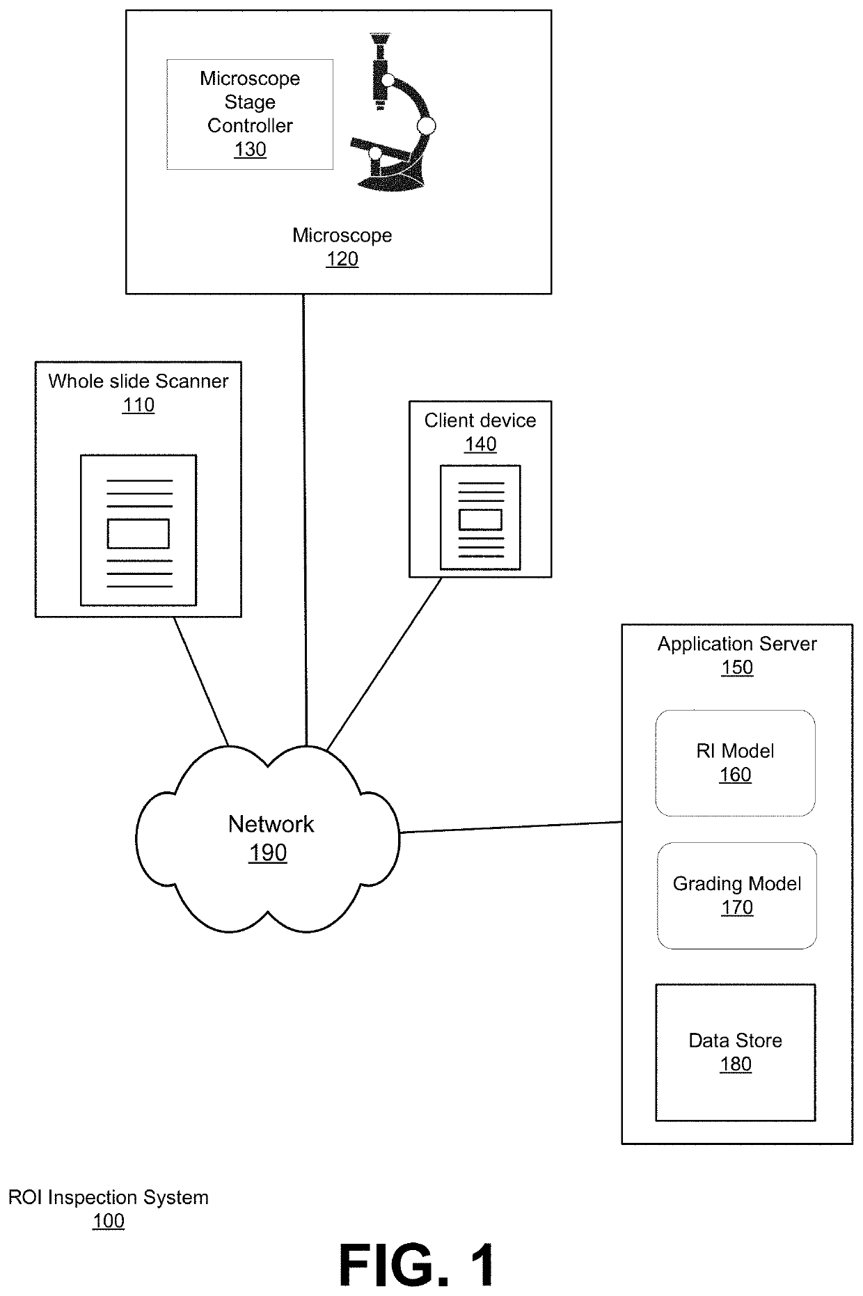
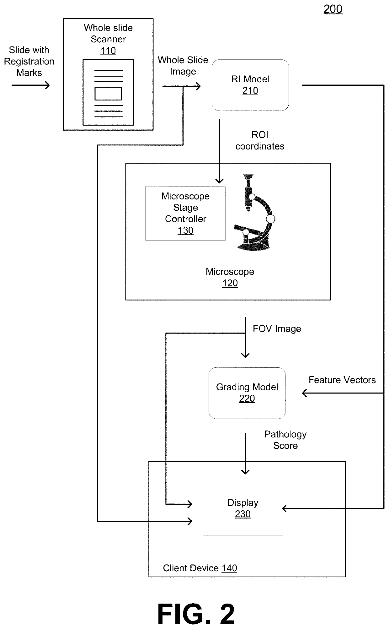
[0037]FIG. 1 shows a region of interest (ROI) inspection system 100 for identifying ROIs on a microscope slide corresponding to regions that are likely to be of interest to a pathologist, and for providing automated control of a microscope and / or augmented reality guidance to assist a pathologist in analyzing the identified ROIs.
[0038]The ROI inspection system 100 includes a whole slide scanner 110, a client computing device 140, and an application server 150 coupled by a network 190. ROI inspection system 100 further includes a microscope 120 with a computer-controlled microscope stage controller 130. The application server 150 comprises a region identification (RI) model 160, a grading model 170, and a data store 180, according to some embodiments. Although FIG. 1 illustrates only a single instance of most of the components of the ROI inspection system 100, in practice more than one of each component may be present, and additional or fewer components may be used...
PUM
 Abstract
Description
Claims
Application Information