Exosomes and uses thereof in diseases of the brain
a brain disease and exosome technology, applied in the field of exosomes, can solve the problems of unfavorable treatment of se, and abnormal electroencephalogram pattern, and achieve the effect of halting or reducing cognitive and memory impairmen
- Summary
- Abstract
- Description
- Claims
- Application Information
AI Technical Summary
Benefits of technology
Problems solved by technology
Method used
Image
Examples
example 1
Preparing the AI Exosome Intranasal Composition, the SE Animal Model
[0059]The procedure described in Kim et al (2016) was employed in the preparation of the A1 exosome preparation. Generally, a population of stem cells, such as mesenchymal stem cells, will be cultured under the conditions defined below, and the cell culture media in which the stem cells were cultured will be collected and screened to select a population of extracellular vesicles (EVs) having a defined set of characteristics.
[0060]Preparations of tissue-derived mesenchymal stem cells (MSCs) may vary in their characteristics depending on many factors, including the properties of the donor of the tissue and the tissue site from which the cells are obtained from the same donor. A preparation of MSCs derived from human bone marrow (defined as Donor 6015) from an NIH-sponsored center for distribution of MSCs was used that met the classical in vitro criteria for MSCs, and ranked among the top three of 13 MSC preparations i...
example 2
l Administration of A1 Exosomes (EVs) Target Hippocampus in an ES In Vivo Model
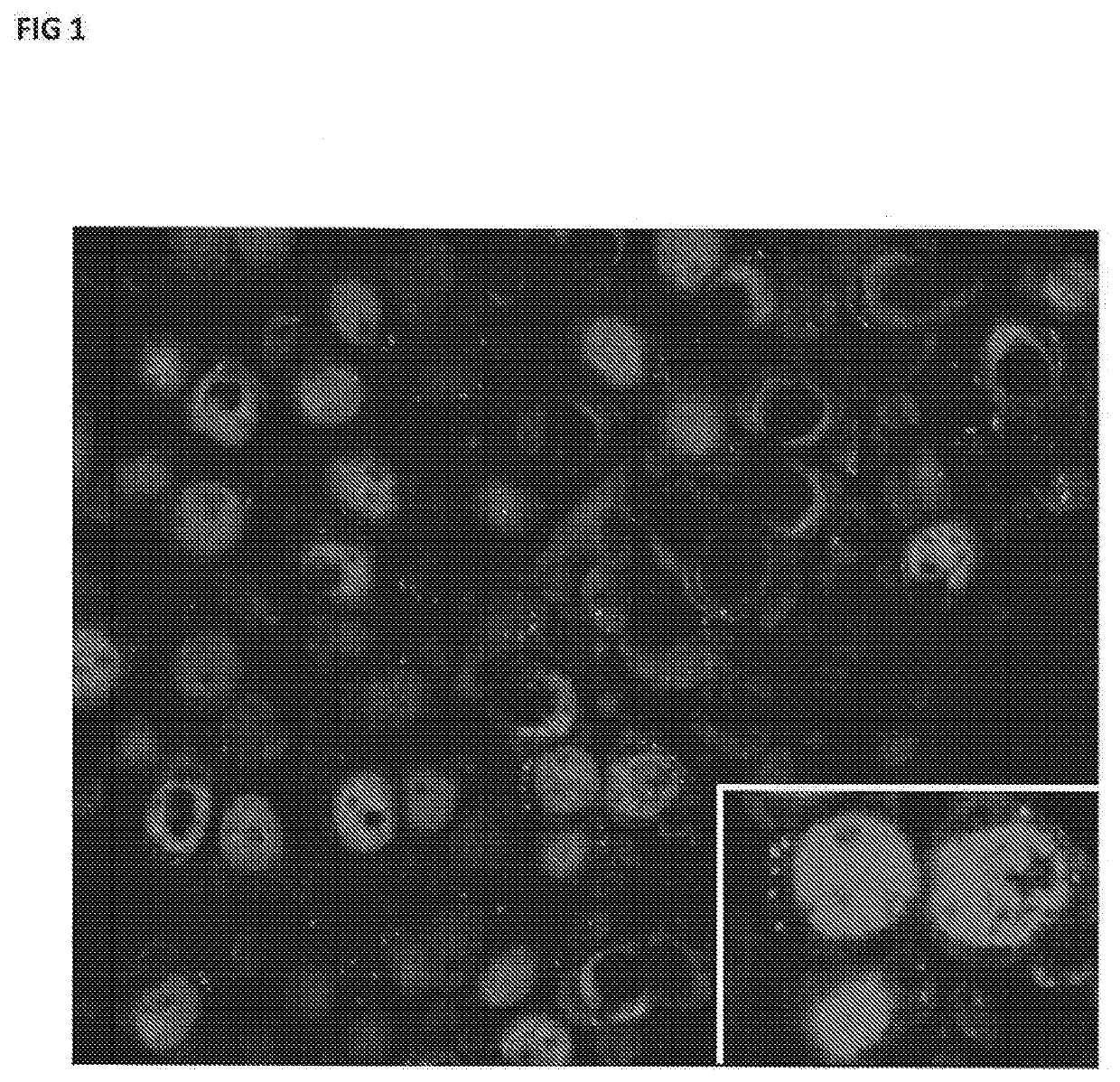
[0097]The present example demonstrates that in vivo intranasal administration of the A1 EV preparation reach the hippocampus (CA3 region) within 6 hours after intranasal administration. FIG. 1 shows distribution of PKH26 labeled EVs in the cytoplasm of CA3 pyramidal neurons of the hippocampus at 6 hours after intranasal administration.
[0098]Thus, intranasal administered EVs target the area of the hippocampus associated with neurodegeneration after SE.
example 3
mmatory Cytokine and Chemokine Levels in the Hippocampus In Vivo
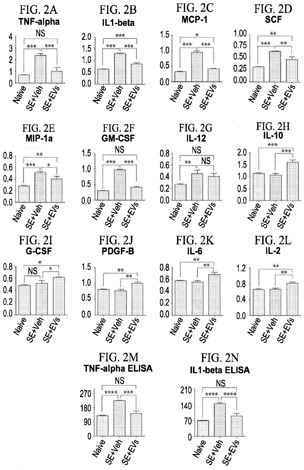
[0099]The results of this study are presented at FIG. 2.
[0100]This example demonstrates that intranasally administered EVs target a tissue, the hippocampus, which is the area of neurodegeneration after SE. This example also shows that this administration of EVs intranasally prevented the up-regulation of 9 different pro-inflammatory proteins (cytokines and chemokines in the hippocampus). These pro-inflammatory proteins include TNFa, IL-1b, MCP-1, MIP-1a, GMCSF, IL-12, SCF, IFNg and IGF-1.
[0101]EV administration intranasally is also shown here to enhance the concentration of anti-inflammatory cytokine IL-10 (see FIG. 2). Thus, intranasal EV treatment after SE significantly extinguishes a major inflammatory response in the hippocampus.
[0102]The above results demonstrate that EV treatment after SE significantly extinguishes a major inflammatory response in the hippocampus.
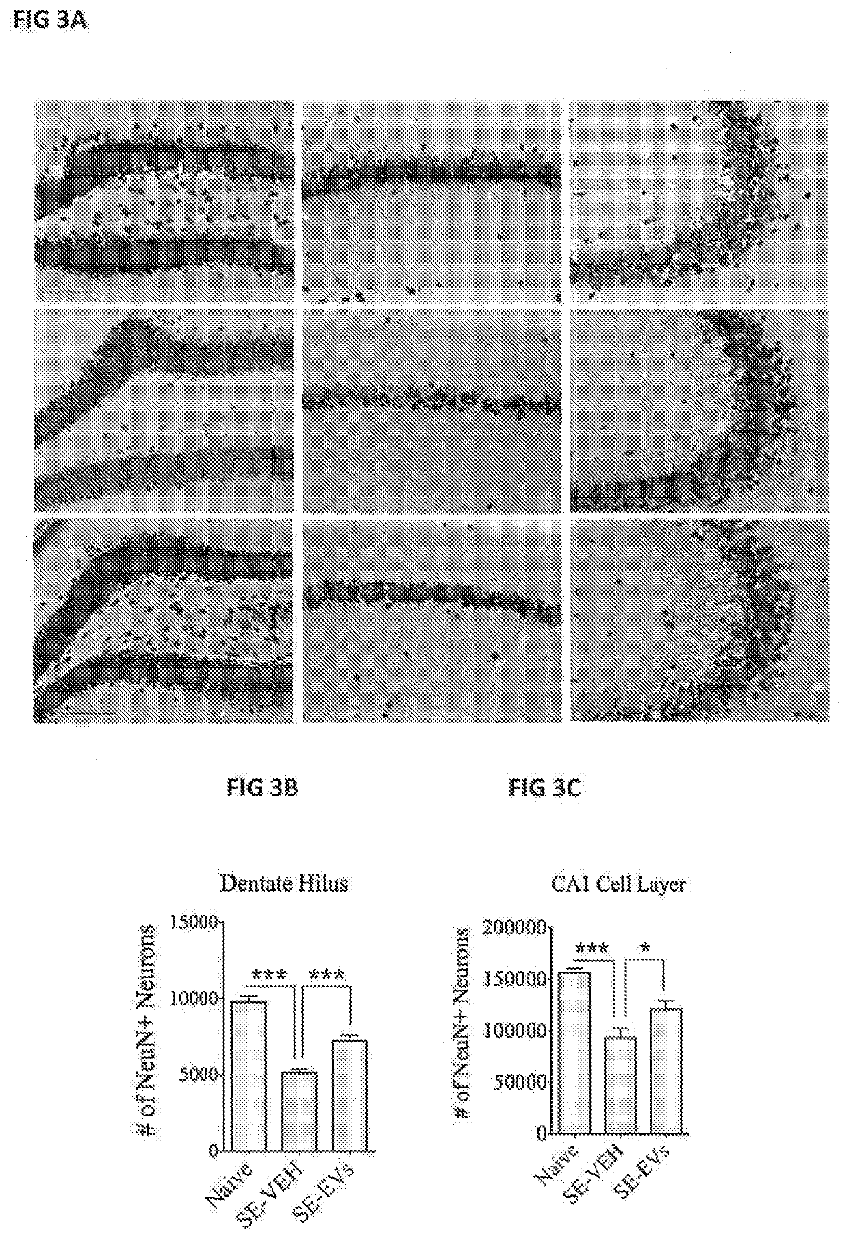
[0103]EV administration is also shown to enhance th...
PUM
 Abstract
Description
Claims
Application Information
 - R&D
- Intellectual Property
- Life Sciences
- Materials
- Tech Scout
- Unparalleled Data Quality
- Higher Quality Content
- 60% Fewer Hallucinations
Browse by: Latest US Patents, China's latest patents, Technical Efficacy Thesaurus, Application Domain, Technology Topic, Popular Technical Reports.
© 2025 PatSnap. All rights reserved.Legal|Privacy policy|Modern Slavery Act Transparency Statement|Sitemap|About US| Contact US: help@patsnap.com


