Medical image processing method and device using machine learning

Pending Publication Date: 2022-07-28
INDAL COOPERATION FOUND CHONBUK NAT UNIV
View PDF0 Cites 0 Cited by
  • Summary
  • Abstract
  • Description
  • Claims
  • Application Information

AI Technical Summary

Benefits of technology

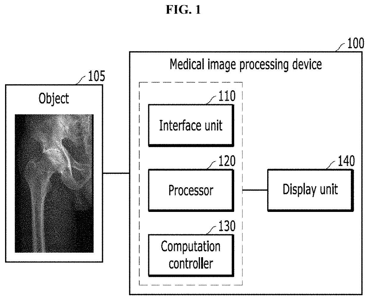
The present patent is about a medical image processing method and device for identifying bones and predicting bone diseases using machine learning. This helps determine which joint to use in surgery. The method also matches color to each bone and displays it to make it easier for surgeons to see. Additionally, the method can predict the shape of damaged hip joints and provide medical support for reconstruction surgery. Overall, this technology makes bone identification and disease prediction easier and more accurate for surgeons.

Problems solved by technology

As described above, only an indirect method that determines the size and shape of the artificial joint to be used in the surgery in reliance on the surgeon's subject determination has been adopted, and there may be a difference between the size / shape of the prepared artificial joint and the actually necessary size / shape in the actual surgery, resulting in low accuracy of the surgery and the prolonged operative time.
To solve the problem, some foreign artificial joint companies provide their own programs to support artificial joint surgeries, but do not publish or open to the public, and the technical levels of the programs are so low that there are many restrictions for surgeons to use.

Method used


Image

  • Medical image processing method and device using machine learning
  • Medical image processing method and device using machine learning
  • Medical image processing method and device using machine learning

Examples


Embodiment Construction

[0024]Hereinafter, embodiments will be described in detail with reference to the accompanying drawings. However, a variety of modification may be made to the embodiments and the scope of protection of the patent application is not limited or restricted by the embodiments. It should be understood that all modifications, equivalents or substitutes to the embodiments are included in the scope of protection.

[0025]The terminology used in an embodiment is for the purpose of describing the present disclosure and is not intended to be limiting of the present disclosure. Unless the context clearly indicates otherwise, the singular forms include the plural forms as well. The term “comprises” or “includes” when used in this specification, specifies the presence of stated features, integers, steps, operations, elements, components or groups thereof, but does not preclude the presence or addition of one or more other features, integers, steps, operations, elements, components or groups thereof.

[...


PUM


Abstract

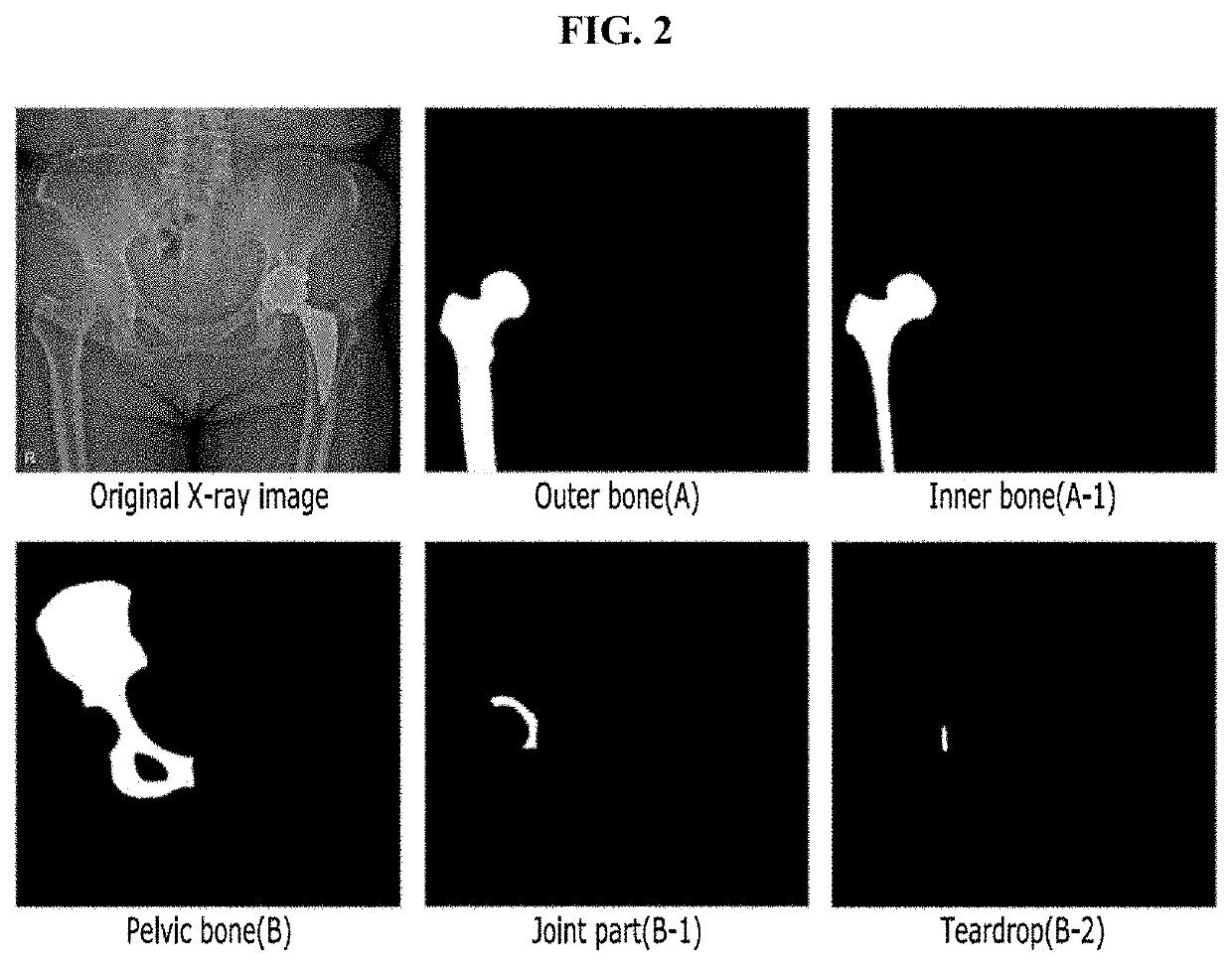
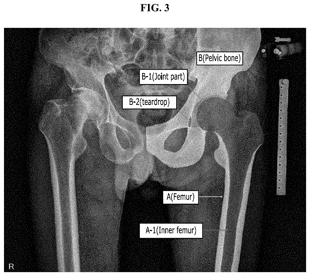
A medical image processing method using machine learning according to an embodiment of the present invention includes acquiring an X-ray image of an object, identifying a plurality of anatomical regions by applying a deep learning technique for each bone structure region that constitutes the X-ray image, predicting a bone disease according to bone quality for each of the plurality of anatomical regions, and determining an artificial joint that replaces the anatomical region in which the bone disease is predicted.

Description

CROSS REFERENCE TO RELATED APPLICATIONS AND CLAIM OF PRIORITY[0001]This application claims benefit under 35 U.S.C. 119(e), 120, 121, or 365(c), and is a National Stage entry from International Application No. PCT / KR2020 / 002866, filed Feb. 28, 2020, which claims priority to the benefit of Korean Patent Application No. 10-2019-0063078 filed in the Korean Intellectual Property Office on May 29, 2019, the entire contents of which are incorporated herein by reference.BACKGROUND1. Technical Field[0002]The present disclosure relates to a medical image processing method and device using machine learning in which human musculoskeletal tissues in a medical image are identified by machine learning and distinguishably displayed in color to determine the size of an artificial joint (implant) that replaces the musculoskeletal tissue more accurately.[0003]In addition, the present disclosure relates to a medical image processing method and device using machine learning in which the diameter and rou...

Claims


Application Information

Patent Timeline
IPC IPC(8): A61B6/00G16H30/40G16H50/20G06T7/00G06V10/25G06V10/774
CPCA61B6/5217A61B6/505A61B6/469G16H30/40G16H50/20G06T2207/30008G06V10/25G06V10/774G06T2207/10116G06T2207/20081G06T7/0014G06V2201/033G06T7/12G06T7/194G06T2207/20084G16H50/50G16H20/40
Inventor YOON, SUN JUNGKIM, MIN WOOOH, II SEOKHAN, KAP SOOKO, MYOUNG HWANCHOI, WOONG
Owner INDAL COOPERATION FOUND CHONBUK NAT UNIV
Why Eureka
Try Eureka