
Method for screening cancer

Active Publication Date: 2015-12-17
NAT DEFENSE MEDICAL CENT
View PDF2 Cites 0 Cited by
  • Summary
  • Abstract
  • Description
  • Claims
  • Application Information

AI Technical Summary

Benefits of technology

The present invention provides a method for screening cervical cancer and other cancers, such as ovarian cancer, liver cancer, colon cancer, breast cancer, oral cancer, endometrial cancer, and sarcoma. This method uses the degree of methylation of specific genes in the in vitro specimen as the screening indicator for the presence or absence of cancer, and has higher sensitivity and specificity compared to existing screening methods such as Pap smear or HPV testing. The method is highly accurate and can help with the determination of abnormal specimens.

Problems solved by technology

However, because it requires physicians to obtain the sample and medical technologists / pathologists to interpret the data from of smear, in addition to high false negative rate and the subsequent delay in diagnosis and treatment of the precancerous lesion, the required quality in human resource and cost is overly high.
For many developing countries, it is difficult to promote.
On the other hand, although HPV testing is highly sensitive, it is also prone to high false positive rate.
Not only the patient worries in vain, but also much medical resource wasted in the follow-up examinations of these false positive patients.
Therefore, there remains a problem of how to elevate the accuracy and convenience of screening methods of cervical cancer for promoting cervical cancer screening.
However, researches in past few years indicate that site-specific hypermethylation (such as in some tumor suppressor genes) is associated with loss of function.
Based on the above, the current methods of cervical cancer screening still have many defects and are not properly designed.

Method used


Image

  • Method for screening cancer
  • Method for screening cancer
  • Method for screening cancer

Examples


example 1

Materials and Methods

1. Materials

[0053]The tested materials comprise a series of intact cervical lesion samples, including: squamous cell carcinoma (SCC), adenocarcinoma (AC), and normal cervical samples. All the cervical samples (SCC+AC, n=20; normal, n=20), ovary samples (cancer of ovary, n=19; normal, n=14), colon samples (Ca of colon, n=18; normal, n=18), liver tissue samples (HCC, n=18; normal, n=18), oral samples (oral Ca, n=20; normal, n=19), endometrial cancer samples (endometrial Ca, n=20; normal, n=20), breast tissue samples (cancer of breast, n=17; normal, n=17), sarcoma samples (sarcoma, n=18; normal, n=16) are obtained from Tri-Service General Hospital in Taipei. The genomic DNA of each sample is extracted using QIAamp DNA kit followed by DNA modification kit (CpGenome™ DNA Modification Kit, Millipore, Temecula, Calif.) produced by Millipore to perform bisulfite modification for analysis of DNA methylation of the whole genome.

2. Analysis of DNA Methylation Using Whole G...

example 2

Screening of Methylated Target Genes

[0061]Screen 14,475 genes using Infinium HumanMethylation27K methylation chips.

[0062](1) Select those having higher methylation rate score (13 value >0.4 and <0.4), combine Gene Expression database (GE07803), and analyze gene enrichment (The Database for Annotation, Visualization and Integrated Discovery, DAVID). 92 genes are selected;

[0063](2) Cancer cell lines are treated with 5-AZc and TSA. Analyze the 92 genes using QRT-PCR to confirm that gene expression is influenced by methylation. 61 genes are left;

[0064](3) Mix the samples to be detected and use MSP to analyze DNA methylation of the 61 genes. 26 genes are left;

[0065](4) Analyze DNA methylation in cancer samples for the 26 genes by MSP. 21 genes are left;

[0066](5) Analyze DNA methylation in cancer tissues for the 21 genes by MSP. 16 genes are left;

[0067](6) Analyze DNA methylation in cancer samples for the 16 genes by Q-MSP. 14 genes are left.

[0068]Confirm the methylation status of genes b...

example 3

Methylation Analysis of Target Genes in Cervical Cancer Samples

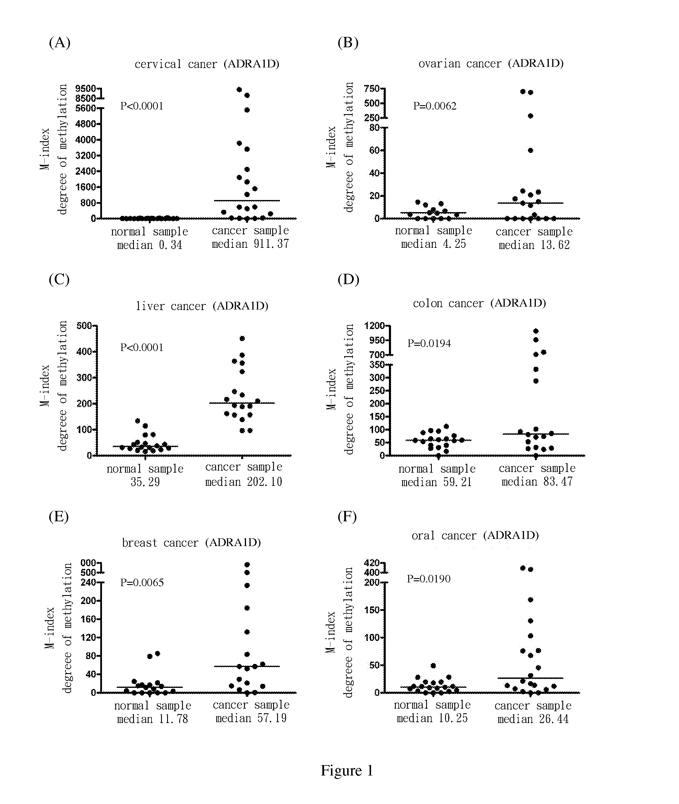
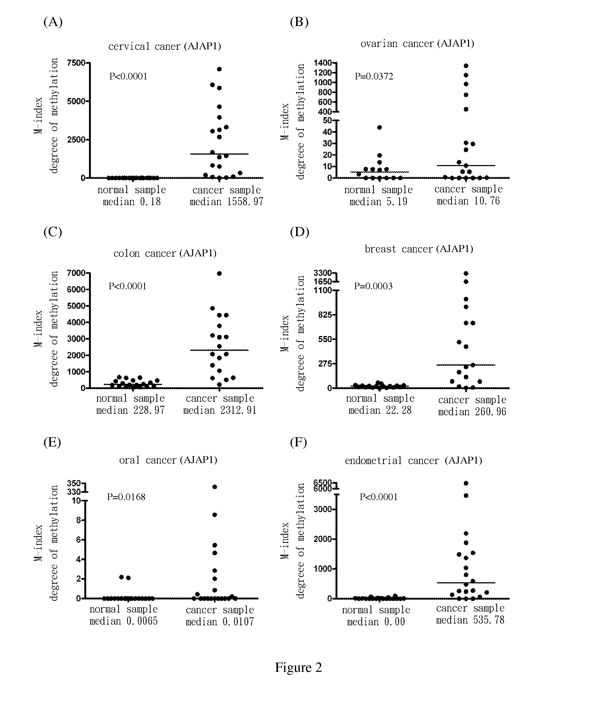
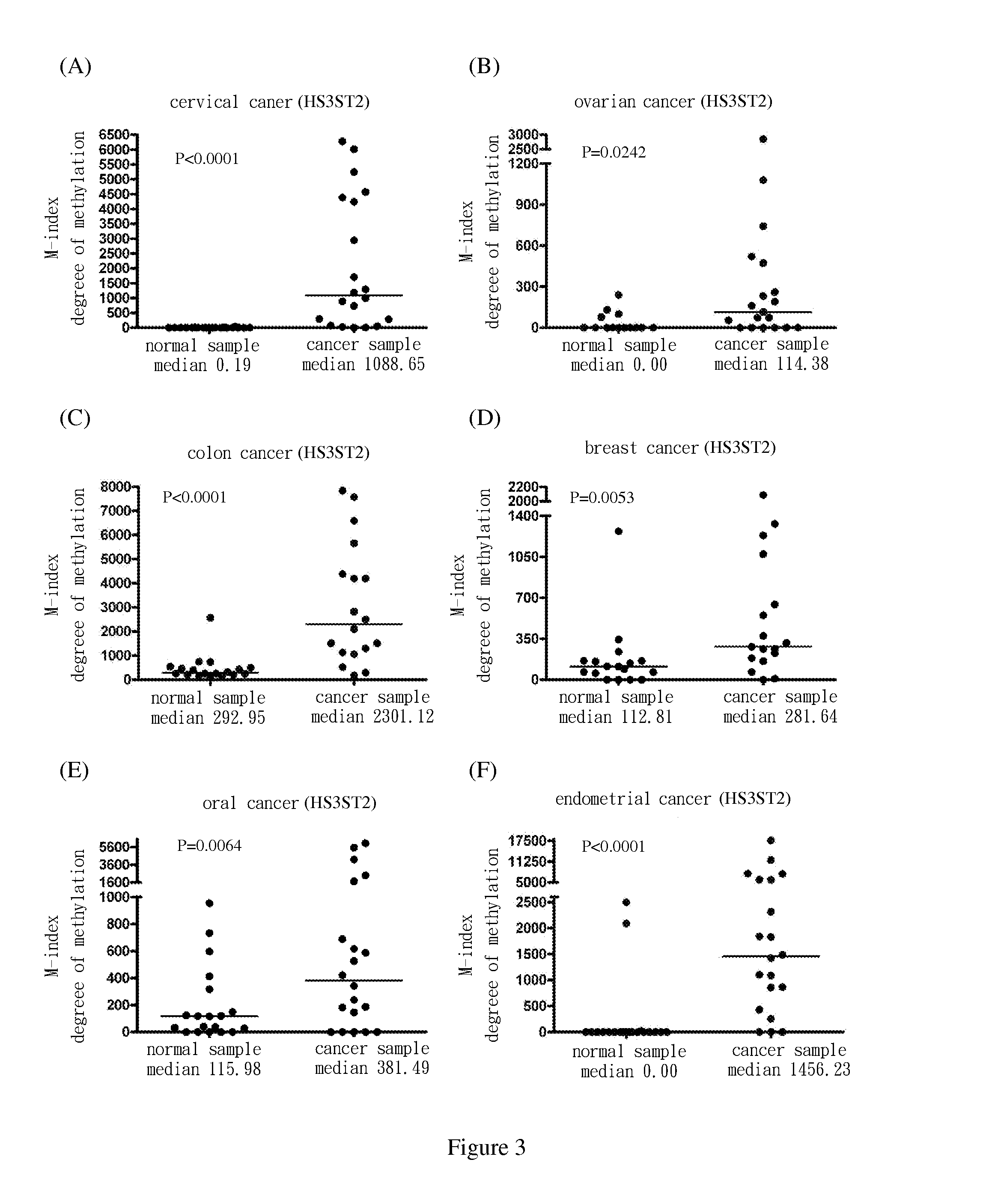
[0069]Use methylation specific PCR (MSP) to analyze the methylation status of the nine target genes in cervical squamous cell cancer samples. The results show the degree of methylation of ADRA1D, AJAP1, HS3ST2, MAGI2, POU4F2, POU4F3, PTGDR, SOX17 and SYT9 in normal cervical samples (SCC N) (the medians are 0.34, 0.18, 0.19, 2.58, 7.62, 0.77, 0.16, 0.17 and 0.31, respectively); and the degree of methylation of ADRA1D, AJAP1, HS3ST2, MAGI2, POU4F2, POU4F3, PTGDR, SOX17 and SYT9 in cervical cancer samples (SCC T) (the medians are 911.37, 1558.97, 1088.65, 713.92, 535.01, 1552.71, 305.84, 248.29 and 551.84, respectively). After Mann-Whitney test, the two groups of data reach P<0.0001 among each group. The difference is statistically significant. The above are as shown in FIG. 1(A), FIG. 2(A), FIG. 3(A), FIG. 4(A), FIG. 5(A), FIG. 6(A), FIG. 7(A), FIG. 8(A), and FIG. 9(A).


PUM

PropertyMeasurementUnit
Fractionaaaaaaaaaa

Abstract

Disclosed in the present invention is a method for screening cancer, comprising the following steps: (1) providing a specimen to be detected; (2) detecting the methylation status of CpG sequence of at least one target gene which is at least one of ADRA1D, AJAP1, HS3ST2, MAGI2, POU4F2, POU4F3, PTGDR, SOX17 and SYT9 in genomic DNA of the specimen; (3) determining whether cancer or precancerous lesions are present in the specimen according to the methylation status of at least one target gene.

Description

FIELD OF THE INVENTION[0001]The present invention relates to method III for screening cancer.BACKGROUND OF THE INVENTION[0002]Cervical cancer is one of the primary causes of death for women worldwide and in Taiwan. According to the statistics by the World Health Organization (WHO) in 2002, cervical cancer is the second cause of death for women cancers worldwide, only subsequent to breast cancer. Periodic cervical cancer screening is the best way of cervical cancer prevention. Currently, there are two methods for cervical cancer screening. One is the most commonly known Pap smear and the other is HPV testing. Pap smear is performed by obtaining the discharge from the cervical, and observing whether cancerous lesion occurs in the detached epithelial cells by microscopy for early-stage detection of cervical cancer. HPV testing is performed by using polymerase chain reaction (RT-PCR) or Hybrid Capture to examine whether human papilloma virus (HPV) is present in the sample.[0003]However,...

Claims


Application Information

Patent Timeline
IPC IPC(8): C12Q1/68
CPCC12Q1/6886C12Q2600/118C12Q2600/112C12Q2600/154
Inventor LAI, HUNGCHENG
Owner NAT DEFENSE MEDICAL CENT
Learn More