High-resolution cerebrospinal fluid-suppressed t2*-weighted magnetic resonance imaging of cortical lesions

a cerebrospinal fluid and magnetic resonance imaging technology, applied in the field of high-resolution, cerebrospinal fluid suppressed t2*-weighted magnetic resonance imaging (mri) of cortical lesions, can solve the problems of obscuring the visualization of cortical lesions, difficult to detect cortical lesions in routine imaging, and difficult to visualize mri from afar, so as to achieve high isotropic resolution, suppress vascular signals, and resolution

Pending Publication Date: 2022-08-25
UNITED STATES OF AMERICA
View PDF6 Cites 0 Cited by
  • Summary
  • Abstract
  • Description
  • Claims
  • Application Information

AI Technical Summary

Benefits of technology

[0009]The T2*-weighted sequence may comprise a 3D-T2*-weighted multi-shot acquisition sequence, and may further comprise a T2-prepared inversion pulse (T2Prep) followed by an inversion pulse. The T2Prep may suppress CSF signals, and the inversion time of the inversion pulse may be tuned to the null point of CSF. The T2Prep may comprise a 90x-180y(4)-90x pulse, and the duration of the T2-Prep pulse may be based on the calculated time at which gray matter (GM) and white matter (WM) signals are equal based on their initial magnetization values. The signal data acquired from the T2*-weighted sequence may be motion corrected before producing the one or more high resolution images. The one or more high resolution images may be processed from magnitude and phase images produced by the T2*-weighted sequence. The T2*-weighted sequence may comprise performing vascular crushing before the acquisition, which may suppress vascular signals. An echo train may be acquired in a centric fashion.
[0010]The method may further comprise performing one or more of a T1-weighted magnetization-prepared 2 rapid acquisition gradient echoes (MP2RAGE) sequence and a fluid-attenuated inversion recovery (FLAIR) sequence. The signal data produced by one or more of MP2RAGE and FLAIR may be used to produce one or more images. The one or more images produced by the T2*-weighted and one or more of MP2RAGE and FLAIR sequences may be indicative of the presence of the cortical lesion. The images produced by the T2*-weighted sequence may be compared to the images produced by the MP2RAGE or FLAIR sequences, or the combination thereof. The comparison may be used to demonstrate the presence or absence of the cortical lesion.
[0011]The T2*-weighted sequence may be repeated 2-4 times, and the signal data acquired from the sequences averaged to produce the one or more high resolution images. In one example, the T2*-weighted sequence may be performed twice. The average may be a voxel-wise median.
[0012]Further provided herein is an MRI system comprising an MRI device and a computer system. The MRI device may comprise a magnet system to apply a polarizing magnetic field about at least a portion of a subject arranged in the MRI system; a plurality of gradient coils to apply a gradient field to the polarizing magnetic field; and, a radiofrequency (RF) system to apply a RF excitation field to a region of interest in the subject and acquire signal data therefrom. The polarizing magnetic field may be 1.5, 3, or 7 tesla. The computer system may have a processor and a memory. The computer system may perform a T2*-weighted sequence by controlling the MRI device, wherein the sequence suppresses CSF signals; acquire high resolution signal data generated by the sequence; and, produce one or more high resolution images from the signal data. The computer system may control the MRI device to sensitize the T2*-weighted sequence with a diffusion gradient; and, produce diffusion-weighted images at a high isotropic resolution of about 0.6 mm or finer, which may be 0.6 mm. The diffusion gradient may suppress CSF signals, and the b-value of the diffusion gradient may be 500-1200 s / mm2 or higher, which may be 500-1200 s / mm2. The computer system may control the MRI device to repeat the T2*-weighted sequence; and, may average the signal data acquired from the T2*-weighted sequences to produce the one or more high resolution images. The average may be a voxel-wise median. The computer system may motion correct the signal data acquired from the T2*-weighted sequence before producing the one or more high resolution images.
[0013]The T2*-weighted system may comprise a 3D-T2*-weighted multi-shot acquisition sequence, and further comprise a T2-prepared inversion pulse (T2Prep) followed by an inversion pulse, wherein the T2Prep suppresses CSF signals. The T2Prep may comprise a 90x-180y(4)-90x pulse, and the duration of the T2-Prep pulse may be based on the calculated time at which GM and WM signals are equal based on their initial magnetization values. The computer system may tune the inversion time of the inversion pulse produced by the MRI device to the null point of CSF. The computer system may control the MRI device to repeat the T2*-weighted sequence 2-4 times, particularly twice; and, average the signal data acquired from the T2*-weighted sequences to produce the one or more high resolution images. The average may be a voxel-wise median. The computer system may motion correct the signal data acquired from the T2*-weighted sequence before producing the one or more high resolution images. The computer system may process magnitude and phase images produced by the T2*-weighted sequence to generate the one or more high resolution images.
[0014]The computer system may control the MRI device to perform vascular crushing before the acquisition to suppress vascular signals. The computer system may acquire an echo train in a centric fashion. The computer system may also control the MRI device to further perform one or more of a MP2RAGE and a FLAIR sequence, acquire MP2RAGE signal data and FLAIR signal data from these one or more sequences to produce one or more MP2RAGE high-resolution images, FLAIR high-resolution images, or a combination thereof. The one or more high-resolution images from the one or more T2*-weighted sequences may be compared to the high-resolution images from the one or more MP2RAGE and FLAIR sequences. The comparison may be indicative of a cortical lesion. In one example, the T2*-weighted sequence is performed twice in combination with the MP2RAGE and FLAIR sequences.

Problems solved by technology

While cortical lesions are known from histopathology studies to be common in MS, they have so far been difficult to visualize on MRI, especially at clinically accessible magnetic field strength.
Cortical lesions are difficult to detect in routine imaging due to the small dynamic range in signal changes induced by the lesions in comparison to the overall dynamic range.
In addition, the close proximity of the cortical lesion, especially of the superficial or “subpial” type of lesion, to cerebrospinal fluid (CSF), can greatly obscure its visualization if the signal intensities from the two tissue types are similar.
Currently available approaches that have been designed to detect cortical lesions at 3 T—including double inversion recovery (DIR) and phase-sensitive inversion recovery (PSIR) imaging—are more sensitive to these signal changes than more standard MRI techniques, but are still poorly sensitive to many cortical lesions, especially subpial lesions, which are thought to underlie substantial clinical disability.
DIR, a sequence with suppressed cerebrospinal fluid (CSF) and white matter signal, is limited by low signal and artifacts, leading to low sensitivity for cortical lesions and high false positive rates.
PSIR has also been used for cortical lesion visualization at 3 T. While PSIR appears to improve sensitivity to leukocortical lesions, data on improvement in subpial lesion detection has been inconsistent.

Method used


Image

  • High-resolution cerebrospinal fluid-suppressed t2*-weighted magnetic resonance imaging of cortical lesions
  • High-resolution cerebrospinal fluid-suppressed t2*-weighted magnetic resonance imaging of cortical lesions
  • High-resolution cerebrospinal fluid-suppressed t2*-weighted magnetic resonance imaging of cortical lesions

Examples


example 1

High-Resolution T2*-Weighted, Cerebrospinal Fluid-Suppressed MR Imaging of Cortical Lesions

[0049]This example demonstrates that two different approaches based on high-resolution T2*-weighted sequences that suppress CSF signals (the DWI approach and the IR-SWIET approach) are capable of detecting cortical lesions better than existing approaches.

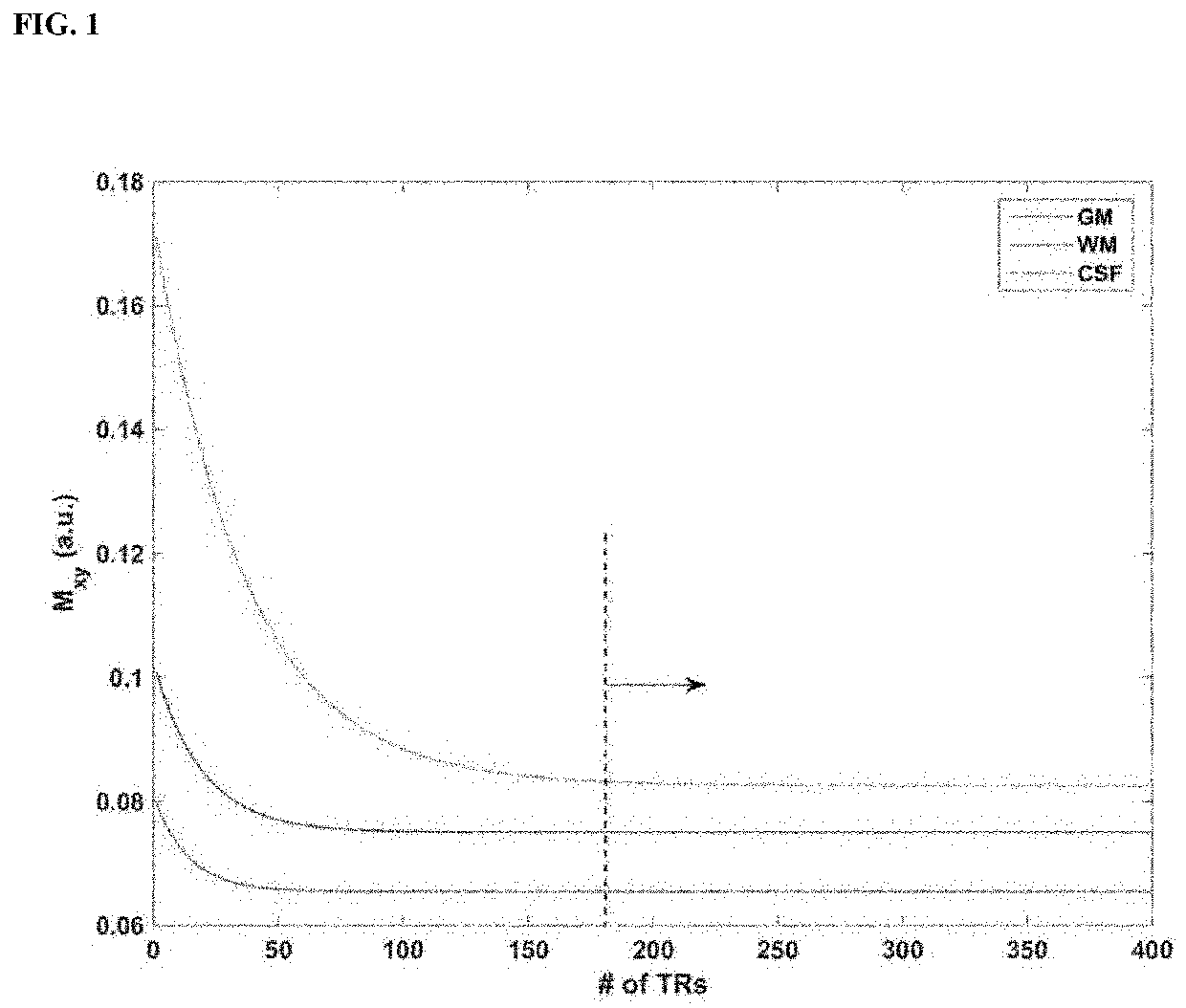
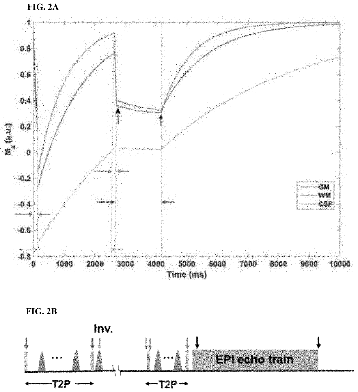
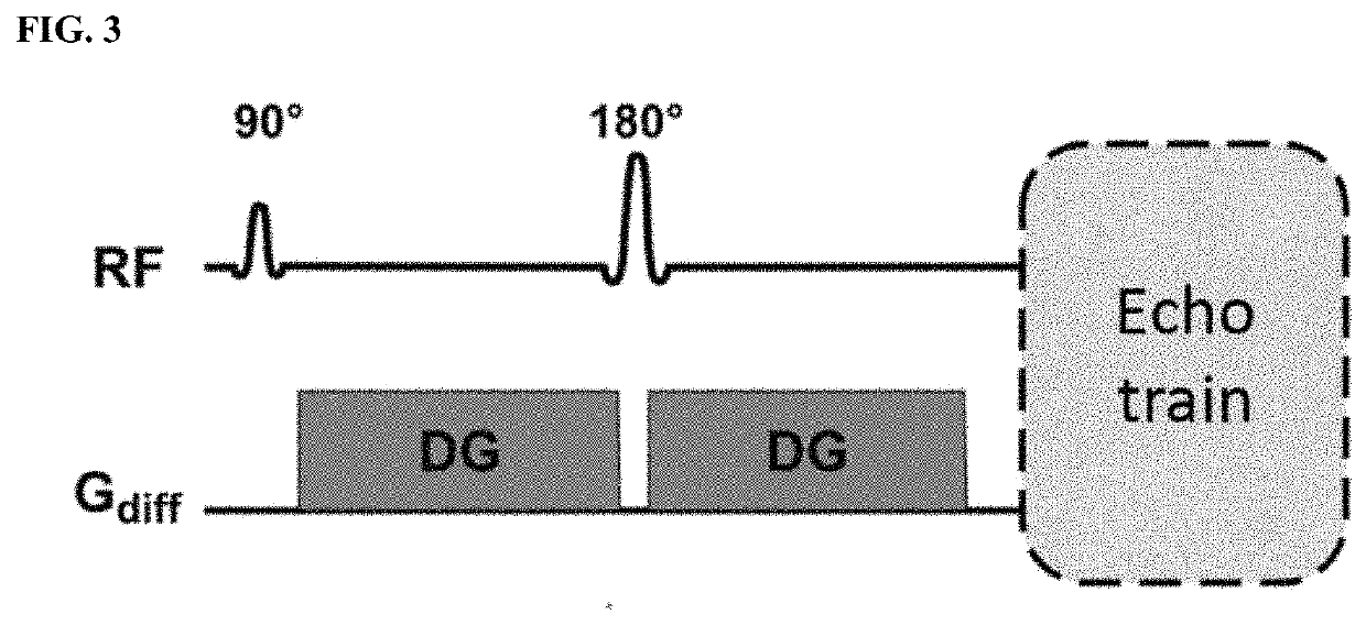
[0050]FIGS. 1-3 show the schematics for the IR-SWIET and DWI approaches.

[0051]FIG. 4 shows that a T2*-weighted sequence that includes inversion pulse with acquisition at null point of CSF and with T2-preparations (IR-SWIET) and the DWI approach improve the visualization of cortical lesions as compared to a T2*-weighted or FLAIR sequence alone.

example 2

Inversion Recovery Susceptibility Weighted Imaging with Enhanced T2 Weighting (IR-SWIET) for Detecting Multiple Sclerosis Lesions

[0052]This example demonstrates the design and evaluation of a 3D sequence (IR-SWIET) that suppresses CSF while maintaining T2* contrast with additional T2 weighting for visualizing cortical and white matter lesions in MS. Multiple sclerosis is an inflammatory, demyelinating, and neurodegenerative disease of the central nervous system. Iron deposition visualized through T2*-weighted imaging has been thought to be a surrogate biomarker for different lesion characteristics (Haacke E M, et al., J Magn Reson Imaging 2009; 29(3):537-544). Iron concentration changes have been observed in newly forming lesions of MS patients (Zhang Y, et al., AJNR Am J Neuroradiol 2016:1629-1635), while central veins visualized by SWI or similar approaches may be a distinguishing feature of MS lesions (Sati P, et al., Nat Rev Neurol 2016:714-722).

[0053]3D interleaved echo planar ...


PUM


Abstract

Provided herein are methods and systems for high-resolution, cerebrospinal fluid-suppressed T2*-weighted magnetic resonance imaging of cortical lesions.

Description

FIELD OF THE INVENTION[0001]The present invention relates to methods and systems for high-resolution, cerebrospinal fluid-suppressed T2*-weighted magnetic resonance imaging (MRI) of cortical lesions.BACKGROUND OF THE INVENTION[0002]Cortical lesions are common in neurologic disease, including multiple sclerosis (MS). Pathological changes in the cerebral cortex in MS include demyelination, neuronal loss, gliosis, and loss of intracellular iron. There is evidence that the pathophysiology of these lesions differs from that of lesions in white matter. Cortical lesions, and especially subpial cortical lesions that touch the pial surface of the brain, may form independently from white matter lesions, perhaps due to leptomeningeal inflammation. In addition, cortical lesions may contribute independently to disability. MRI is an essential tool for clinical management of and research on MS and other neurological conditions, including diagnosis, prognosis, response to treatment, and outcome mea...

Claims


Application Information

Patent Timeline
Patent Type & Authority Applications(United States)
IPC IPC(8): G01R33/56G01R33/563G01R33/565A61B5/00A61B5/055
CPCG01R33/5607G01R33/5602G01R33/56341G01R33/56509G01R33/5608A61B5/0042A61B5/055A61B5/4064A61B5/4082A61B5/7264A61B2576/026A61B5/743
Inventor REICH, DANIEL SALOBECK, ERIN SAVNERNAIR, GOVINDGAI, NEVILLE DALI
Owner UNITED STATES OF AMERICA
Why Eureka
Try Eureka