Percutaneous heart valve delivery systems

a delivery system and heart valve technology, applied in the field of percutaneous delivery of heart valves, can solve the problems of differences in contrast, resolution, and x-ray use, and achieve the effects of reducing the risk of heart valve failure, and reducing the safety of patients

Pending Publication Date: 2022-09-22
RGT UNIV OF CALIFORNIA
View PDF0 Cites 0 Cited by
  • Summary
  • Abstract
  • Description
  • Claims
  • Application Information

AI Technical Summary

Problems solved by technology

However, only X-Ray can be used during the procedure for image guidance.
X-Ray is not sufficient for visualization because it is a 2D projection of 3D anatomy that depends on the orientation angle of visualization.
However, differences in contrast, resolution, and artifacts can produce differing results.
However, most of the current technologies are limited by instant deployment, and once the valve is deployed, repositioning and / or percutaneous retrieval is not possible—or at least difficult or potentially problematic.
Placement of the stented valve in a position that is too high (or proximal) can totally or partially obstruct the coronary ostia in a case of aortic implantation, which may result in myocardial infarction or ischemia.
Additionally, if the valve is placed too high in the aorta, it may embolize into the aorta causing significant paravalvular regurgitation.
On the other hand, implantation in a position that is too low (or distal) is accompanied by compression of the atrioventricular (AV) node in the membranous septum, which leads to conduction abnormalities.

Method used


Image

  • Percutaneous heart valve delivery systems
  • Percutaneous heart valve delivery systems
  • Percutaneous heart valve delivery systems

Examples


Embodiment Construction

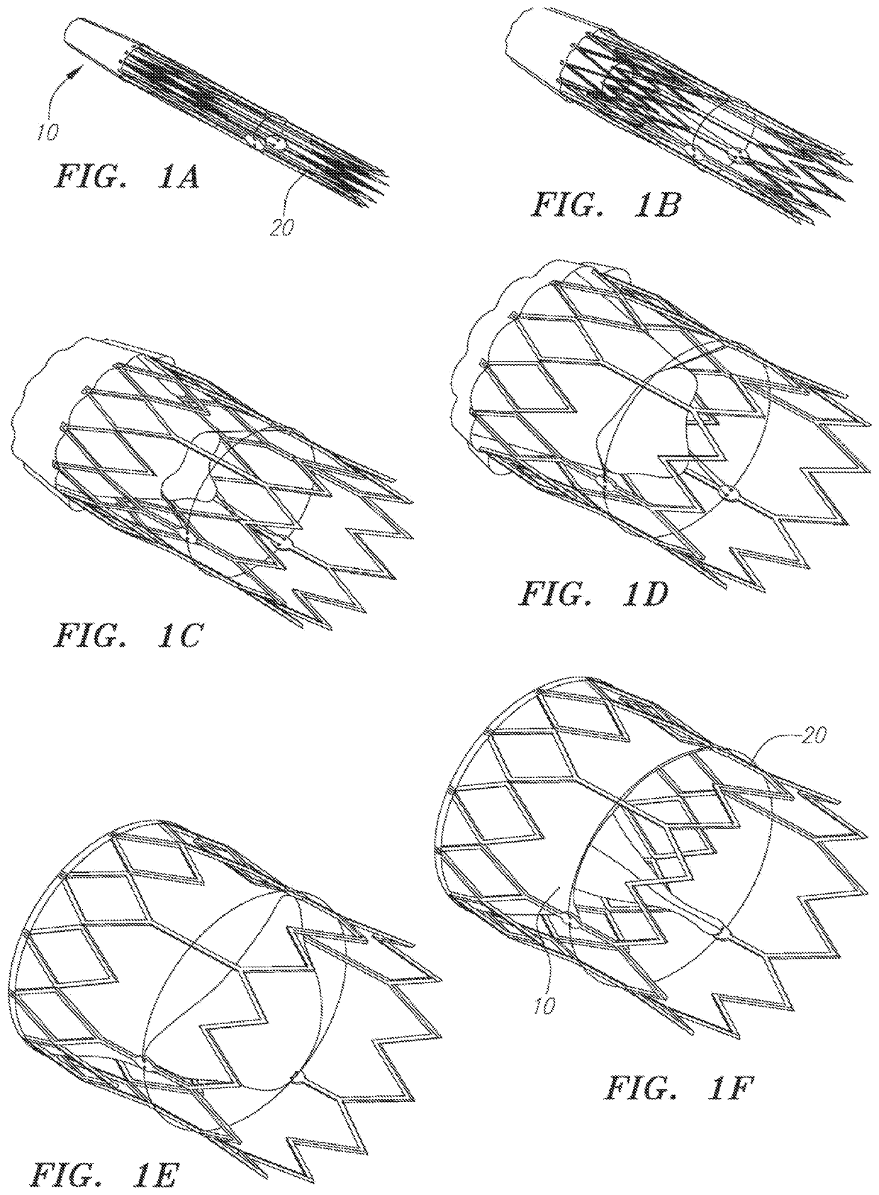
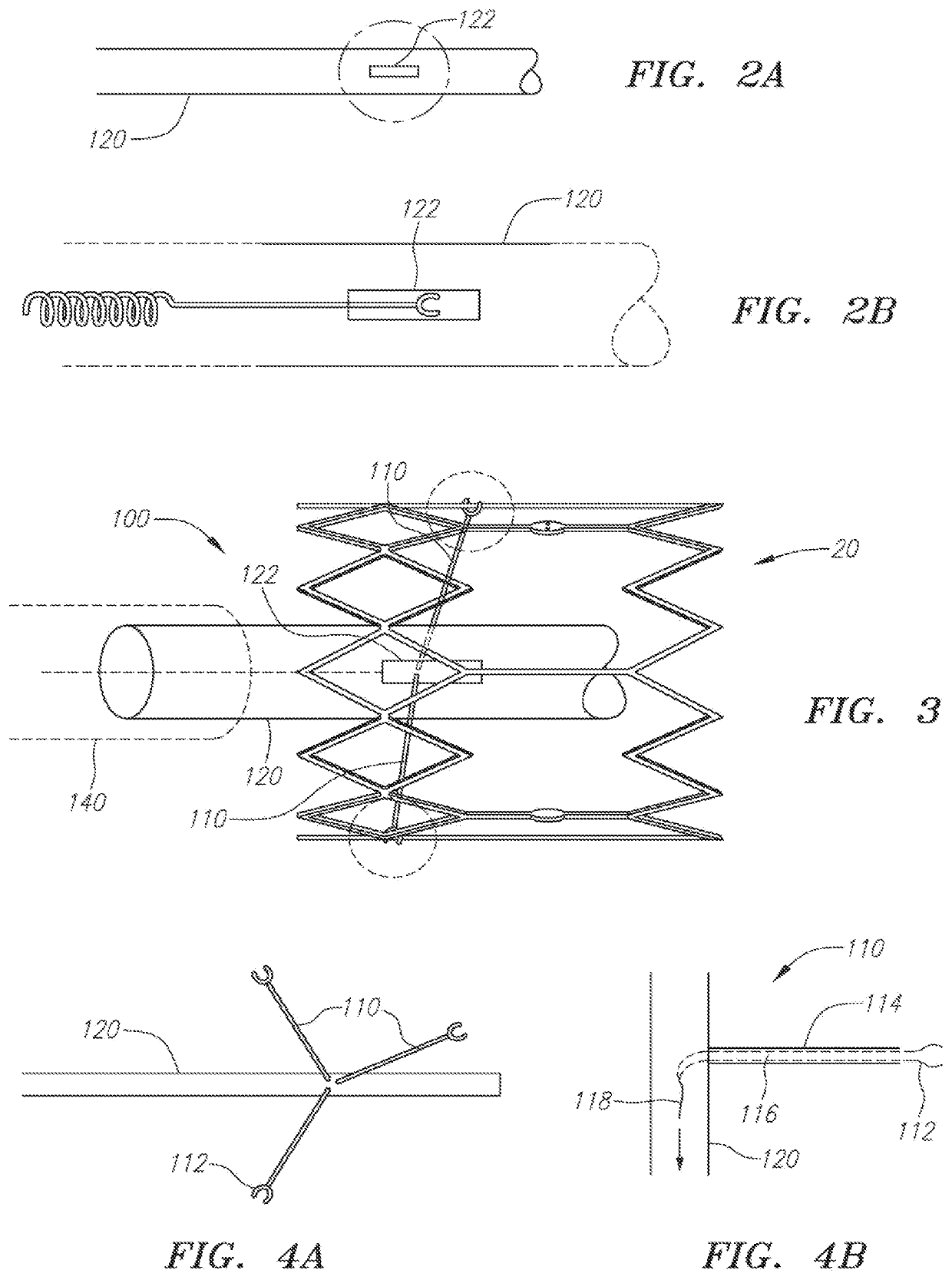
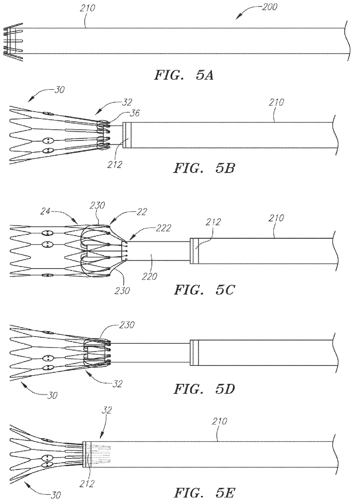
[0051]Various example embodiments are described below. Reference is made to these examples in a non-limiting sense, as it should be noted that they are provided to illustrate more broadly applicable aspects of the devices, systems and methods. Various changes may be made to these embodiments and equivalents may be substituted without departing from the true spirit and scope of the various embodiments. In addition, many modifications may be made to adapt a particular situation, material, composition of matter, process, process act, or step to the objective(s), spirit, or scope of the present inventive subject matter. All such modifications are intended to be within the scope of the claims made herein.

[0052]The present invention features a delivery system (400) comprising an implantable heart valve (401), a valve delivery catheter (410) comprising a first lumen (415), and an ultrasound imaging catheter (420) inserted through the center of the first lumen (415) of the valve delivery ca...


PUM


Abstract

Embodiments described herein address the need for improved catheter devices for delivery, repositioning and/or percutaneous retrieval of the percutaneously implanted heart valves. One embodiment employs a plurality of spring-loaded arms releasably engaged with a stent frame for controlling expansion for valve deployment. Another embodiment employs a plurality of filaments passing through a distal end of a pusher sleeve and apertures in a self-expandable stent frame to control its state of deployment. With additional features, lateral positioning of the stent frame may also be controlled. Yet another embodiment includes plurality of outwardly biased arms held to complimentary stent frame features by overlying sheath segments. Still another embodiment integrates a visualization system in the subject delivery system. Variations on hardware and methods associated with the use of these embodiments are contemplated in addition to those shown and described

Description

CROSS-REFERENCES TO RELATED APPLICATIONS[0001]This application is a continuation-in-part, and claims benefit of U.S. patent application Ser. No. 14 / 221,194 filed Mar. 20, 2014, which is a continuation of U.S. patent application Ser. No. 13 / 862,302, filed Apr. 12, 2013, which claims the benefit of and priority to U.S. Provisional Patent Application No. 61 / 732,117, filed Nov. 30, 2012, U.S. Provisional Patent Application No. 61 / 682,663, filed Aug. 13, 2012, and U.S. Provisional Patent Application No. 61 / 623,410, filed Apr. 12, 2012.[0002]This application is also a continuation-in-part, and claims benefit of U.S. patent application Ser. No. 14 / 221,194 filed Mar. 20, 2014, which is a continuation-in-part of PCT / US12 / 49645, filed Aug. 3, 2012, which claims the benefit of and priority to U.S. Provisional Patent Application No. 61 / 515,679, filed Aug. 5, 2011, and U.S. Provisional Patent Application No. 61 / 666,657, filed Jun. 29, 2012. All preceding applications are incorporated by referenc...

Claims


Application Information

Patent Timeline
Patent Type & Authority Applications(United States)
IPC IPC(8): A61F2/24A61B8/08A61B90/00A61B8/12
CPCA61F2/2439A61F2/2427A61B8/0841A61B8/0891A61B90/37A61B8/12A61B2090/3782A61B8/0883A61F2/2436A61F2/9522A61F2240/007A61F2/2496
Inventor KHERADVAR, ARASHSU, JIMMY L.
Owner RGT UNIV OF CALIFORNIA
Why Eureka
Try Eureka