Synthetic four-dimensional computed tomographic image generation

a computed tomographic image and synthetic technology, applied in the field of magnetic resonance imaging, can solve problems such as blurred structure boundaries

Pending Publication Date: 2022-09-22
KONINKLJIJKE PHILIPS NV
View PDF0 Cites 0 Cited by
  • Summary
  • Abstract
  • Description
  • Claims
  • Application Information

AI Technical Summary

Problems solved by technology

33 that the misregistration of multi-echo Dixon image can con...

Method used


Image

  • Synthetic four-dimensional computed tomographic image generation
  • Synthetic four-dimensional computed tomographic image generation
  • Synthetic four-dimensional computed tomographic image generation

Examples


Embodiment Construction

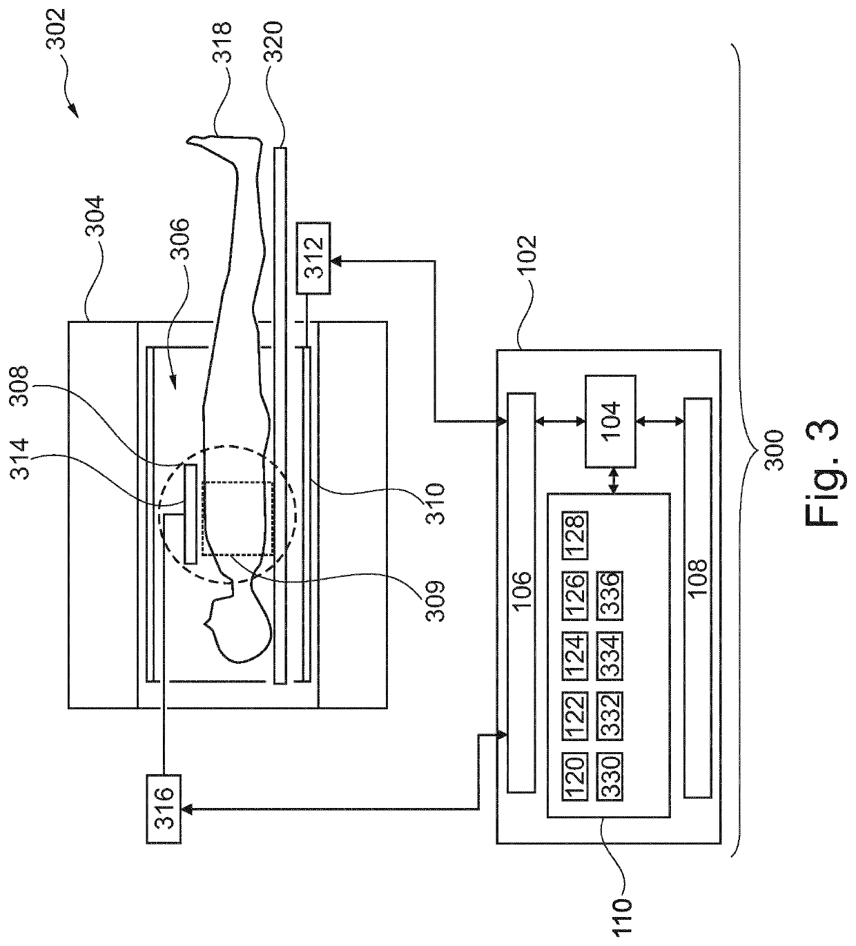
[0066]Like numbered elements in these figures are either equivalent elements or perform the same function. Elements which have been discussed previously will not necessarily be discussed in later figures if the function is equivalent.

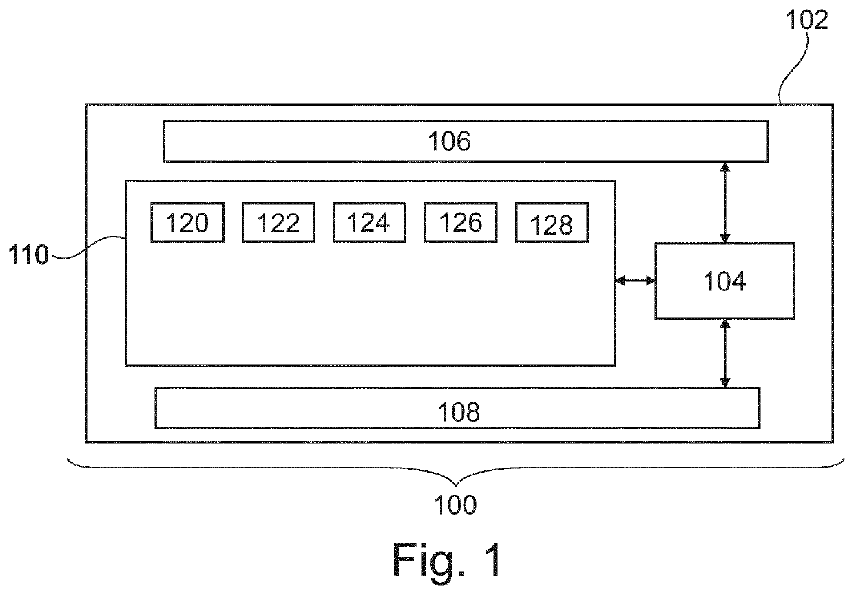
[0067]FIG. 1 illustrates an example of a medical system 100. The medical system 100 is shown as comprising a computer 102. The computer 102 comprises a processor 104. The processor 104 is intended to represent one or more processors with one or more processing cores. The processor 104 could for example be distributed amongst multiple computer systems. The processor 104 is connected to an optional hardware interface. The hardware interface 106 may for example be a network interface that enables the processor 104 to communicate with and / or control additional components of the medical system 100. The processor 104 is further shown as being connected to a user interface 108. The user interface 108 may for example optionally comprise a display, user interfac...


PUM


Abstract

Disclosed herein is a medical system (100, 300, 500) comprising a processor (104) configured for controlling the medical system and a memory (110) for storing machine executable instructions. Execution of the instructions causes the processor to receive (200) four-dimensional Dixon magnetic resonance image data (122). The four-dimensional Dixon magnetic resonance imaging data is T1 weighted. The four-dimensional Dixon magnetic resonance image data is synchronized to a respiratory signal (124). Execution of the instructions further causes the processor to reconstruct (202) synthetic four-dimensional computed tomographic image data (12) from the four-dimensional Dixon magnetic resonance imaging data. The four-dimensional Dixon magnetic resonance imaging data is synchronized to the respiratory signal.

Description

FIELD OF THE INVENTION[0001]The invention relates to Magnetic Resonance Imaging, in particular to generation of synthetic computed tomographic images from magnetic resonance images.BACKGROUND OF THE INVENTION[0002]Computer tomography (CT) images are useful for radiotherapy planning. CT image measure the three-dimensional absorption of X-rays by the body. The images can be expressed in terms of a Hounsfield units or as a spatial dependent electron density map. One of these values are typically used by radiotherapy planning software to estimate the absorption of ionizing radiation for radiotherapy. A difficulty in using CT for radiotherapy planning is firstly that it exposes the subject to additional radiation. Another difficulty is that CT is excellent in imaging hard or dense tissues such as bone, but it does not differentiate between different type of soft tissue as well as magnetic resonance imaging (MRI). Commonly during radiation therapy planning both a CT image and an MRI image...

Claims


Application Information

Patent Timeline
IPC IPC(8): G01R33/56G01R33/48G01R33/567A61N5/10A61B5/00A61B5/055
CPCG01R33/5608G01R33/4812G01R33/4828G01R33/567A61N5/1037A61B5/0036A61B5/055A61N2005/1055A61N5/1049A61N5/1068A61N5/1039
Inventor KADBI, MOHAMMAD HOSSEINWARNER, LIZETTEKRUISKAMP, MARINUS JOHAN
Owner KONINKLJIJKE PHILIPS NV
Why Eureka
Try Eureka