Ultrasound endoscope

a technology of ultrasonic endoscopy and endoscopy, which is applied in the field of ultrasonic endoscopy, can solve the problems of difficult monitoring of the position of the puncture needle's fore end within the view field, difficult to accommodate the puncture needle within the length of the inflexible rigid distal end, and difficult to check

Inactive Publication Date: 2000-11-21
FUJI PHOTO OPTICAL CO LTD
View PDF13 Cites 124 Cited by
  • Summary
  • Abstract
  • Description
  • Claims
  • Application Information

AI Technical Summary

Benefits of technology

It is another object of the present invention to provide an ultrasound endoscope which can guarantee safe operation of a puncture instrument with a sharp-pointed puncture needle at the time of protrusion out of an instrument passage or similar treating instrument guide means provided on the endoscope.

Problems solved by technology

It follows that, in case the puncture needle has an increased length, it might become difficult to accommodate the puncture needle within the length of the inflexible rigid distal end section of the endoscopic insertion instrument.
When the angle section of the endoscopic insertion instrument is flexed to turn the rigid distal end section into the direction of a target, however, the puncture needle could be accidentally projected out of the biopsy channel of the endoscopic insertion instrument, depending upon the turning angle.
In this regard, according to the prior art, it has been difficult to monitor the position of the sharp-pointed fore end of the puncture needle within the view field of an endoscopic image pickup until the puncture instrument is projected over a certain length from the distal opening of the endoscopic biopsy channel.
Namely, it has thus far been difficult to check, within the view field of the endoscopic image pickup, the position of the sharp-pointed end of the puncture instrument in or in the vicinity of the distal opening of the biopsy channel.
Therefore, even if the sharp-pointed end of the puncture needle is slightly projected from the distal opening of the biopsy channel for some reason, in many cases this cannot be recognized by the operator.
However, because of a short axial length of the rigid distal end section of the endoscopic insertion instrument, it is often found difficult to retract the rigid puncture needle smoothly into and out of the endoscopic biopsy channel unless it is reduced in length to such an extent as would make deep penetrations infeasible.

Method used


Image

  • Ultrasound endoscope
  • Ultrasound endoscope
  • Ultrasound endoscope

Examples


Embodiment Construction

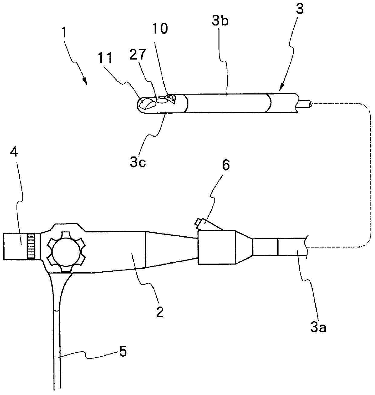
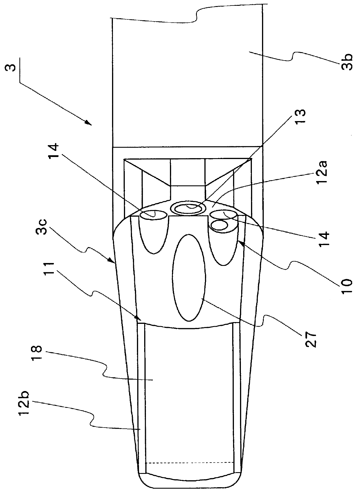
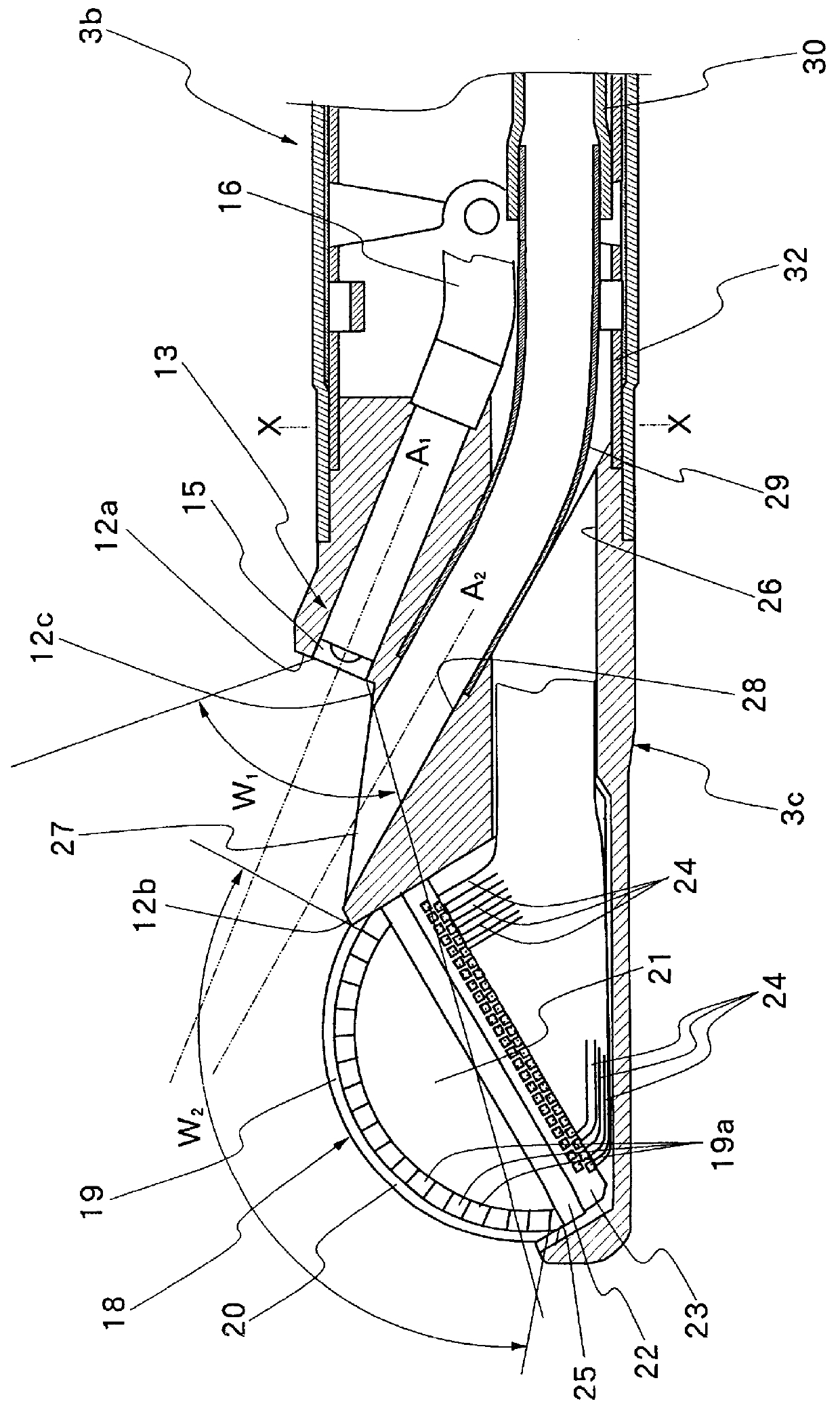
Hereafter, the present invention is described more particularly by way of its preferred embodiment with reference to the accompanying drawings. Firstly, referring to FIG. 1 which shows the general layout of an ultrasound endoscope, indicated at 1 is the ultrasound endoscope, including a head grip assembly 2 to be gripped and manipulated by an operator, and an elongated insertion instrument 3 extended forward from the head grip assembly 2 for introduction into a body cavity of a patient. The endoscopic insertion instrument 3 includes, from its proximal end which is connected to the head grip assembly 2, a flexible section 3a which constitutes a major part of the elongated insertion instrument 3 and which is flexibly bendable in arbitrary directions to comply with bends in a path of insertion, if any, an angle section or flexible joint 3b which is connected to the fore end of the flexible section 3a, and a rigid distal end section 3c constituted by a rigid distal end casing structure ...


PUM


Abstract

An ultrasound endoscope having an endoscopic observation system along with an ultrasound scan system. An outlet opening of an instrument passage, which is shunted from a biopsy channel of the endoscope, is located within a view field of an endoscopic image pickup window, which largely overlaps a scan range of the ultrasound scan system. A puncture instrument having a sharp-pointed rigid needle at the instrument outlet opening, which is in the view field of the endoscopic image pickup, can be monitored by way of the endoscopic observation system to check its position for safety purposes, from a slightly projected position to a largely projected position whenever it spontaneously gets out of the instrument outlet opening. Once the puncture needle is driven into an intracavitary wall toward a diseased portion in target, it can be monitored through the ultrasound scan system.

Description

1. Field of the ArtThis invention relates generally to an ultrasound endoscope to be introduced into a body cavity of a patient for making ultrasound scans of internal body tissues in addition to an endoscopic examination, and more particularly to an ultrasound endoscope which is so arranged as to guarantee safe use of a puncture instrument or similar treating instruments.2. Prior ArtEndoscopes and ultrasound probes are in wide use especially in medical fields for examining intracavitary portions for diagnostic or therapeutical purposes. An endoscopes is used mainly for examination of surface conditions of intracavitary walls, while an ultrasound probe is used for scanning internal body tissues. Besides, there have also been in wide use the so-called ultrasound endoscopes which are equipped with an ultrasound scanning system along with an endoscopic observation system. More particularly, ultrasound endoscopes are generally provided with an endoscopic or optical observation system, i...

Claims


Application Information

Patent Timeline
Patent Type & Authority Patents(United States)
IPC IPC(8): A61B8/12A61B1/00
CPCA61B8/12A61B8/4488A61B8/445
Inventor TANAKA, TOSHIZUMI
Owner FUJI PHOTO OPTICAL CO LTD
Why Eureka
Try Eureka