Method of magnetic resonance imaging of a sample with ex vivo polarization of an MR imaging agent

a magnetic resonance imaging and sample technology, applied in the field of magnetic resonance imaging, can solve the problems of toxic and inability to be administered, polarization is quickly lost, and it takes an unacceptably long time for the medium to reach thermodynamic equilibrium

Inactive Publication Date: 2001-08-21
OXFORD INSTR MOLECULAR BIOTOOLS
View PDF5 Cites 89 Cited by
  • Summary
  • Abstract
  • Description
  • Claims
  • Application Information

AI Technical Summary

Problems solved by technology

After polarisation it may be dissolved in heated water or saline or melted and removed or separated from the OMRI contrast agent where the latter may be toxic and cannot be administered.
Hydrogenation may be conveniently but not necessarily performed in aqueous media and appropriate catalysts for this use should operate efficiently i

Method used


Image

  • Method of magnetic resonance imaging of a sample with ex vivo polarization of an MR imaging agent
  • Method of magnetic resonance imaging of a sample with ex vivo polarization of an MR imaging agent
  • Method of magnetic resonance imaging of a sample with ex vivo polarization of an MR imaging agent

Examples


example 2

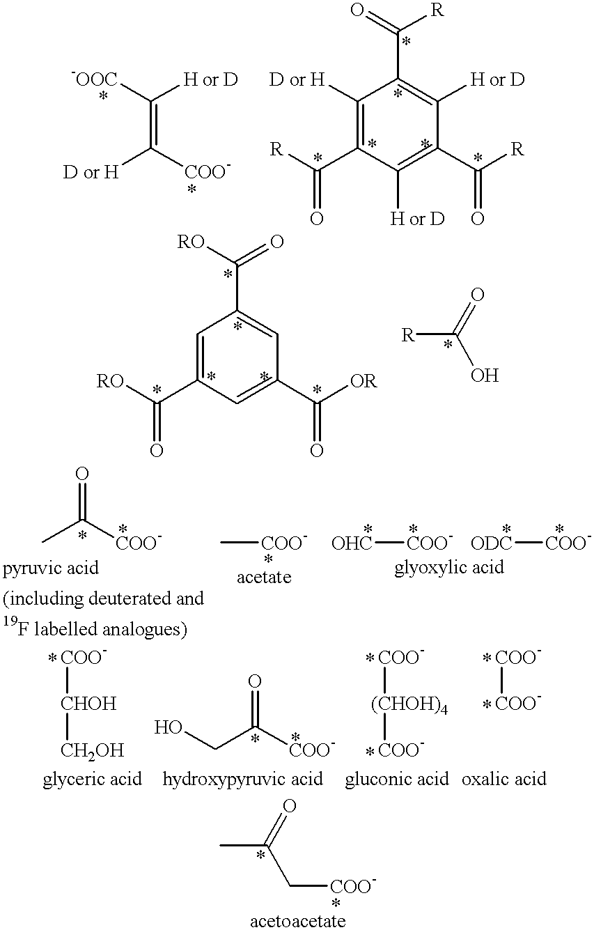
300 mg of sterile Na.sub.2.sup.13 CO.sub.3 or NaH.sup.13 CO.sub.3 is placed inside a 10 ml plastic injection syringe. The gas inside the syringe is enriched with >20% oxygen. The syringe is placed inside a magnet (1-20 T) at a temperature of about 4 K (0.001-5 K) until thermodynamic equilibrium is reached.

The syringe is removed and transported to the subject located in the MRI magnet. 10 ml of sterile Ringers Solution (at 37.degree. C., pH 7.4) is aspirated and injected at a rate of 10 ml / sec immediately after the high T.sub.1 agent has dissolved. .sup.13 C MRI is performed using a fast pulse sequence. T.sub.1 in the blood is about 20 s and the distribution of the agent is followed on the MR imager.

example 3

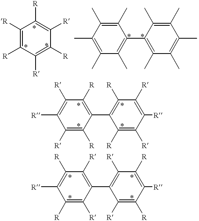
To a sample of sodium acetate (1-.sup.13 C) is added .alpha., .gamma.-bisphenyl-.beta.-phenylallyl benzene complex (5% w / w). The compounds are milled together to give an intimate mixture, which is transferred to a borosilicate glass ampule. This is then repeatedly evacuated and filled with helium. The last time a pressure of a 200 mbar of helium is left in the ampule, which is then flame sealed.

The sample is polarized by microwaves (70 GHz) for at least one hour at a field of 2.5 T at a temperature of 4.2 K. The progress of the polarization process is followed by in situ NMR (fast adiabatic passage). When a suitable level of polarization has been reached, the ampule is rapidly removed from the polarizer and, while handled in a magnetic field of no less than 50 mT, cracked open and the contents are quickly discharged and dissolved in warm (40.degree. C.) water.

experiment 1

n is quickly transferred to a spectrometer and .sup.13 C spectrum with enhanced intensity is recorded.


PUM


Abstract

The present invention provides a method of magnetic resonance investigation of a sample, preferably of a human or non-human animal body, said method comprising the step of ex vivo polarisation of a high T1 agent and wherein the polarising agent is optionally seperated from the high T1 agent before the high T1 agent is administered to the sample.

Description

FIELD OF THE INVNENTIONThis invention relates to a method of magnetic resonance imaging (MRI).BACKGROUND OF THE INVNENTIONMagnetic resonance imaging (MRI) is a diagnostic technique that has become particularly attractive to physicians as it is non-invasive and does not involve exposing the patient under study to potentially harmful radiation such as X-rays.In order to achieve effective contrast between MR images of the different tissue types in a subject, it has long been known to administer to the subject MR contrast agents (e.g. paramagnetic metal species) which effect relaxation times of the MR imaging nuclei in the zones in which they are administered or at which they aggregate. Contrast enhancement has also been achieved by utilising the "Overhauser effect" in which an esr transition in an administered paramagnetic species (hereinafter an OMRI contrast agent) is coupled to the nuclear spin system of the imaging nuclei. The Overhauser effect (also known as dynamic nuclear polari...

Claims


Application Information

Patent Timeline
IPC IPC(8): A61K49/18A61K49/08A61K49/06G01R33/28A61B5/055A61B6/00A61K49/00G01N24/08G01R33/48G01R33/62
CPCA61K49/08A61K49/10A61K49/18G01R33/282G01R33/5601A61K49/1815G01R33/62
Inventor ARDENKJ.AE BUTTED.R-LARSON, JAN HENRIKAXELSSON, OSKARGOLMAN, KLAESHANSSON, GEORGLEUNBACH, IBPETERSSON, STEFANWISTRAND, LARS-GORAN
Owner OXFORD INSTR MOLECULAR BIOTOOLS
Why Eureka
Try Eureka