Immunoassay of IGF family of peptides, their binding proteins and related molecules in dried whole blood filter paper spots

a technology of igf family and antibody, which is applied in the field of immunoassay of igf family of peptides, their binding proteins and related molecules in dried whole blood filter paper spots, can solve the problems of controversial hormone status and problematic blood sampling through venipunctur

Inactive Publication Date: 2002-02-19
BECKMAN COULTER INC
View PDF12 Cites 18 Cited by
  • Summary
  • Abstract
  • Description
  • Claims
  • Application Information

AI Technical Summary

Problems solved by technology

The clinical assessment of growth hormone status has been controversial, primarily due to the episodic nature of GH secretion, its relatively short circulating half-life and considerable variability in GH measurement by different assay methods (Lee P D K, et al.
In children blood sampling by way of venipuncture has been problematic.

Method used


Image

  • Immunoassay of IGF family of peptides, their binding proteins and related molecules in dried whole blood filter paper spots
  • Immunoassay of IGF family of peptides, their binding proteins and related molecules in dried whole blood filter paper spots
  • Immunoassay of IGF family of peptides, their binding proteins and related molecules in dried whole blood filter paper spots

Examples


example 2

Extraction of IGF-I

Determination of IGFs require their dissociation from IGFBPs. Procedures for direct measurement of IGFs by sample acidification to a pH of about 2.0 to dissociate the complexes followed by neutralization to slightly alkaline pH prior to analysis have been developed by DSL. Thus, the optimized IGF-I extraction protocol combined a pre-extraction step with sample acidification and neutralization steps using the corresponding reagents developed for direct analysis of the IGFs by DSL. Two hundred .mu.L of the IGF pre-extraction buffer (see below) was added to each well containing a single unknown or control dried blood disc and incubate shaken at room temperature for 1 h to allow complete elution of IGF from the filter paper. To the extract was then added 200 .mu.L of an acidification buffer, containing 0.4 M glycine-HCl, pH 2.0 incubated as above for 30 min, and neutralized by adding 400 .mu.L of a neutralizaiton buffer, containing 0.85 M Tris and 0.1% sodium dodecyls...

example 3

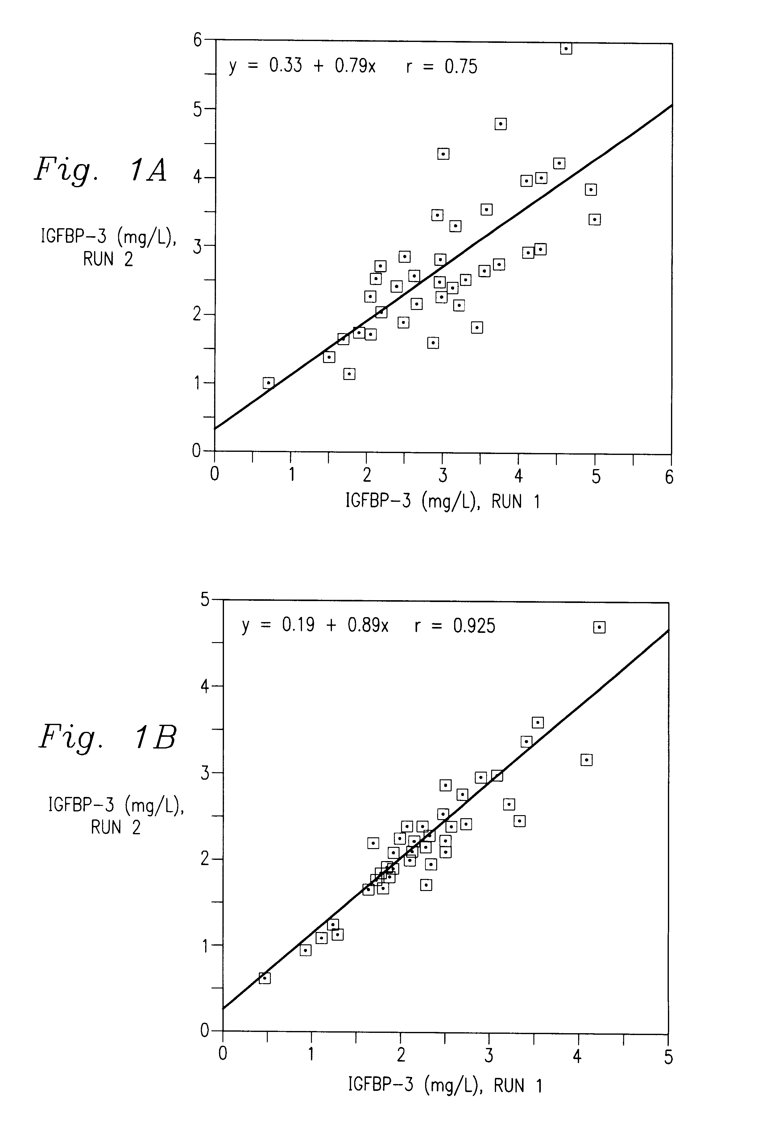
Extraction of IGFBP-3

For the optimized IGFBP-3 extraction, 0.5 mL of the IGFBP-3 extraction buffer (see below) was added to each well containing a single unknown or control dried blood disc. IGFBP-3 was eluted by incubating the wells shaken for 1 h at room temperature. The extract was used for IGFBP-3 determination as well as determination of IGFBP-2 which is similarly released into the surrounding media. The same extraction procedure was used for elution of IGFBP-1 from dried blood filter paper spots, except that discs were extracted with 0.25 mL of the IGFBP-3 extraction buffer.

example 4

Optimization of the Extraction Procedures

Extraction efficiency of IGF-I and IGFBP-3 was investigated using a number of extraction media and extraction time (30-120 min). The extraction media used were as follows: (1), deionized water (dH.sub.2 O); (2), IGF-I pre-extraction buffer (0.005 mol / L Tris, pH 7.0, 0.5 mL / L Tween-20); (3), IGFBP-3 extraction buffer (0.05 mol / L sodium borate, pH 8.5, 9 g / L NaCl, 10 g / L bovine serum albumin (BSA), 0.1 g / L thimerosal); (4) 0.05 mol / L sodium phosphate, pH 7.4, 0.9 g / L NaCl, 10 g / L BSA, 1 mL / L Tween-20, 0.1 g / L thimerosal.

To explore the possibility of using a common extraction procedure and reagents for IGF-I and IGFBP-3, dried blood spot discs were also directly extracted by the addition of 0.5 mL of the IGF acidification buffer / well followed by 1 h incubation as above and addition of 0.5 mL of the IGF neutralization buffer and mixing. The performance of the direct acidification protocol for IGFand IGFBP-3 analysis was then evaluated.

Both IGF-I ...


PUM

PropertyMeasurementUnit
timeaaaaaaaaaa
pHaaaaaaaaaa
diameteraaaaaaaaaa

Abstract

The present invention relates to screening or testing for insulin like growth factors, insulin like growth factor binding proteins and / or acid labile subunit by the use of a solid support. Blood is collected onto a solid support, such as paper, and subsequently the analytes of interest are extracted for testing.

Description

1. Field of the InventionThe present invention relates to screening or testing for insulin like growth factors and their binding proteins and related molecules ("IGF / IGFBPs"). More specifically the present invention relates to a method for screening IGF / IGFBPs from whole blood spotted on a solid support. Additionally, the present invention relates to a test kit for screening or testing IGF / IGFBPs on the solid support.2. Description of the Prior ArtInsulin-like growth factors (IGFs) belong to a family of peptides that share a high degree of structural homology with insulin. IGFs (IGF-I and IGF-II) have important mitogenic and anabolic actions that are mediated by their binding to specific high affinity cell surface receptors (Jones J I, et al. Endocrin Rev 1995; 16:3-34). IGF-I and IGF-II are produced in multiple tissues and are present in blood and other biological fluids in association with specific high affinity IGF binding proteins (IGFBPs). Six structurally homologous IGFBPs wit...

Claims


Application Information

Patent Timeline
Patent Type & Authority Patents(United States)
IPC IPC(8): G01N33/74
CPCG01N33/74Y10T428/24802G01N2333/65G01N2333/4745
Inventor KHOSRAVI, M. JAVADMISTRY, JEHANGIRDIAMANDI, ANASTASIA
Owner BECKMAN COULTER INC
Why Eureka
Try Eureka