
Safety cryotherapy catheter

a safe and cryotherapy technology, applied in the direction of catheters, cooling devices, lighting and heating devices, etc., can solve the problem of undesirable cell necrosis

Inactive Publication Date: 2003-02-04
BOSTON SCI SCIMED INC
View PDF50 Cites 259 Cited by
  • Summary
  • Abstract
  • Description
  • Claims
  • Application Information

AI Technical Summary

Benefits of technology

Inhibition of excessive cell growth is desirable when such treatments are employed so as to reduce and / or eliminate any associated hyperplasia and to maintain the patency of a body lumen. The present invention allows for cryotherapy treatment of a target portion within the body lumen of a patient in a very controlled and safe manner, particularly when using fluid capable of cooling tissues below a target temperature range.
Treatment according to this first aspect of the present invention can be effected by positioning the first balloon within the blood vessel adjacent a target portion. The "target portion" will often be a length within the blood vessel which is at risk of hyperplasia, typically as a result of balloon angioplasty (or some other treatment). Cryogenic cooling fluid is introduced into the first balloon (in which it often vaporizes) and exhausted. The second balloon expands to radially engage the vessel wall. The target portion is cooled to a temperature which is sufficiently low for a time which is sufficiently long to inhibit excessive cell proliferation. Heat transfer will be inhibited between the first and second balloons by the thermal barrier so as to limit cooling of the target portion. The inhibited cooling treatment will be directed at all or a portion of a circumferential surface of the body lumen, and will preferably result in cell growth inhibition, but not necessarily in significant cell necrosis. Particularly in the treatment of arteries before, during, and / or following balloon angioplasty, cell necrosis may be undesirable if it increases the hyperplastic response. Thus, the present invention will cool target tissue to a limited cooling temperatures to slow or stop cell proliferation.
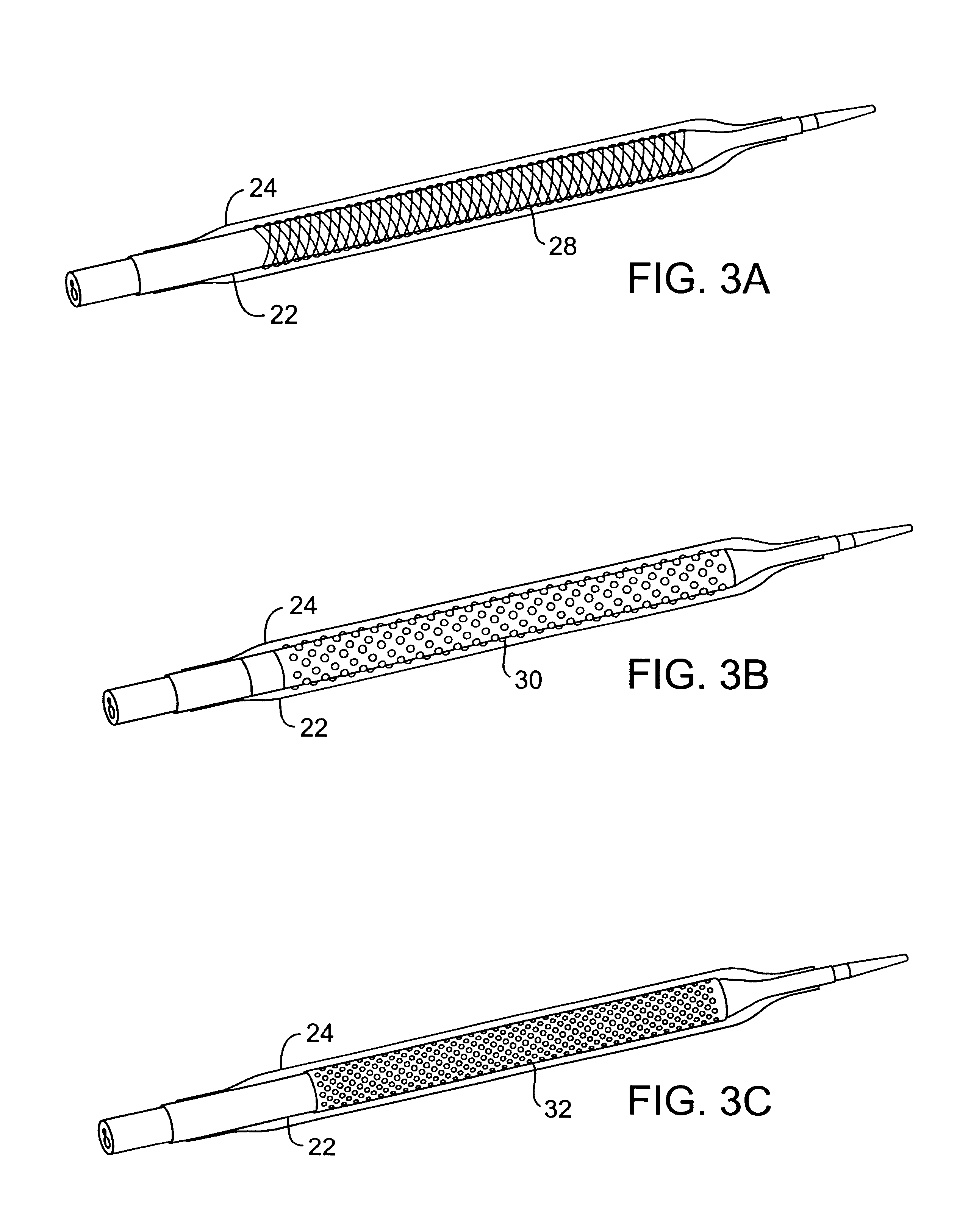
The thermal barrier may comprise a gap maintained between the balloons by a filament. The filament typically comprises a helically wound, braided, woven, or knotted monofilament. The thermal barrier may also comprise a gap maintained between the balloons by a plurality of bumps on an outer surface of the first balloon or an inner surface of the second balloon. Alternatively, the thermal barrier may comprise a sleeve. The sleeve can be solid or perforated. The catheter of the present invention may also be equipped with a guidewire lumen that extends axially outside the exhaust lumen to minimize the occurrence of cryogenic fluid entering the blood stream via the guidewire lumen.
Advantageously, the cryotherapy system can monitor the integrity of both balloons during cooling to ensure that no cryogenic fluid is escaping from the first balloon or blood entering from the second balloon. Further, in the event of a failure, the fluid shutoff can prevent the delivery of additional cryogenic fluid into the supply lumen while the second balloon acts to contain any cryogenic fluid that may have escaped the first balloon.
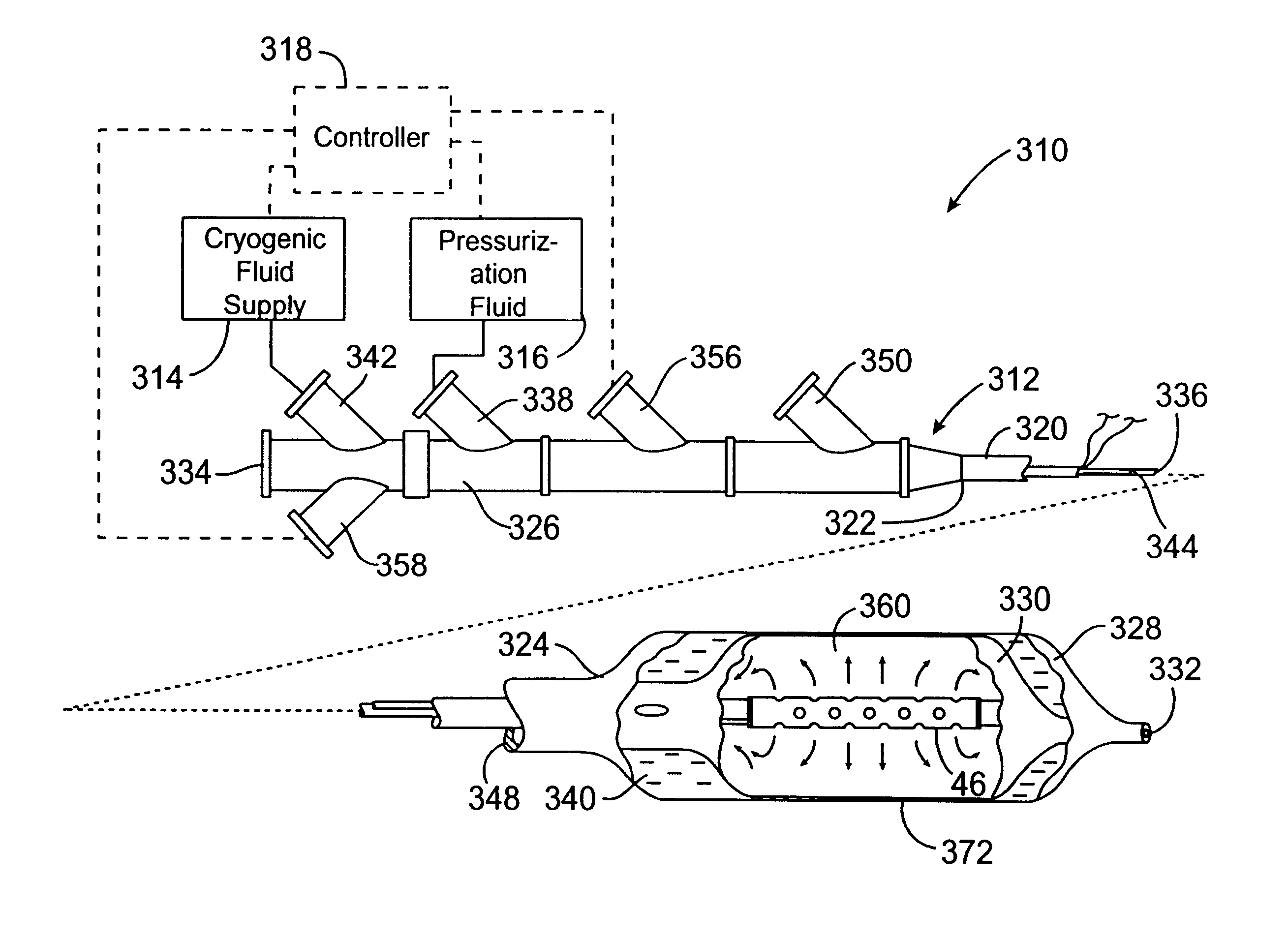
The nebulizer may comprise at least one port in fluid communication with a liquid supply lumen and a gas supply lumen. The liquid supply lumen may further be coaxial with the gas supply lumen. Thus, the nebulizer can introduce a liquid and gas mixture into the first balloon so that pressure and the enthalpy of vaporization of a safe cryogenic fluid within the balloon surface can be independently selected and / or controlled. This in turn allows for improved temperature control of the cryogenic fluid.

Problems solved by technology

Particularly in the treatment of arteries before, during, and / or following balloon angioplasty, cell necrosis may be undesirable if it increases the hyperplastic response.

Method used


Image

  • Safety cryotherapy catheter
  • Safety cryotherapy catheter
  • Safety cryotherapy catheter

Examples


Embodiment Construction

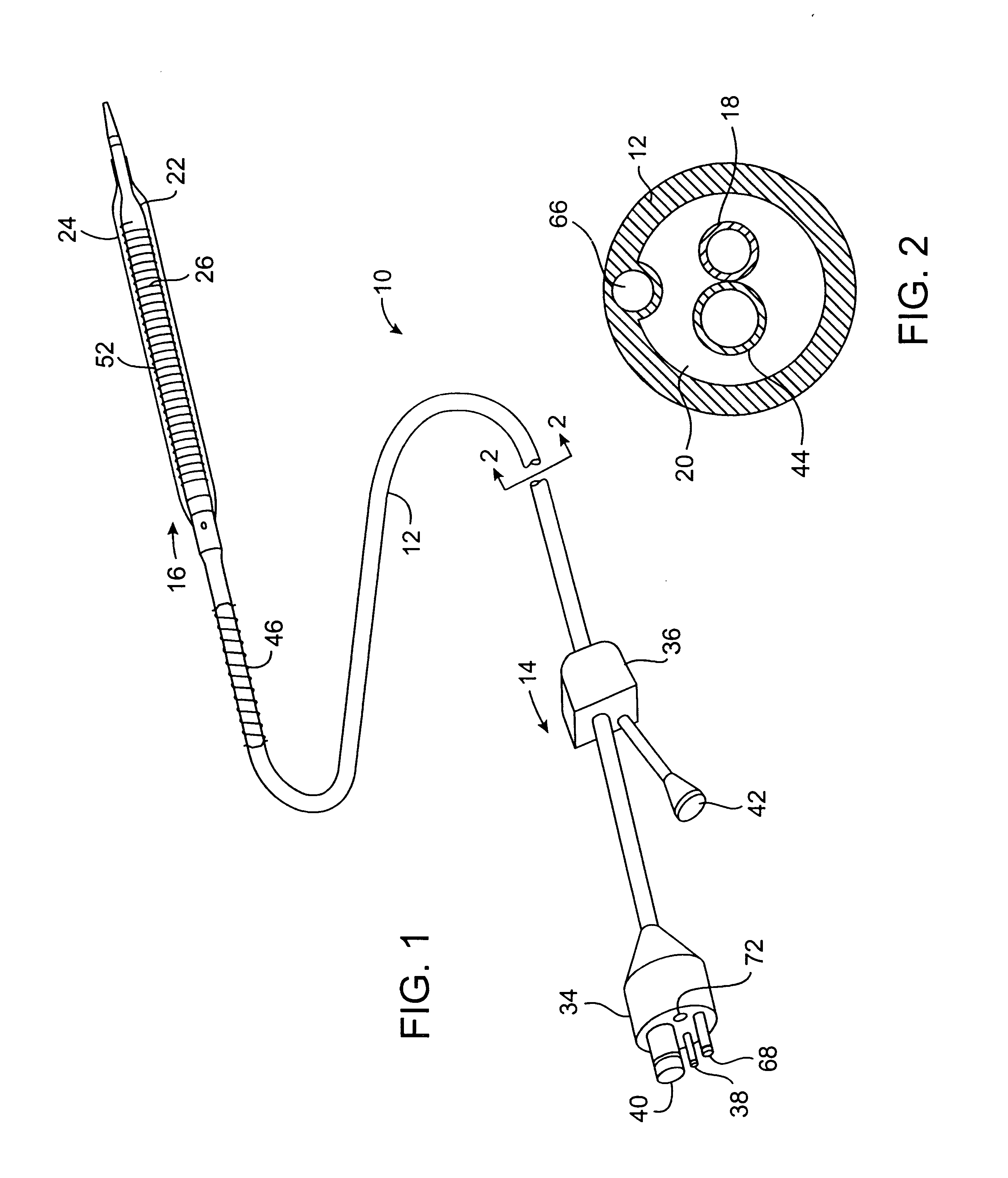
The present invention provides improved cryotherapy devices, systems, and methods for inhibiting hyperplasia in blood vessels. An exemplary cryotherapy catheter 10 constructed in accordance with the principles of the present invention is illustrated in FIGS. 1 and 2. The catheter 10 comprises a catheter body 12 having a proximal end 14 and a distal end 16 with a cooling fluid supply lumen 18 and an exhaust lumen 20 extending therebetween. A first balloon 22 is disposed near the distal end of the catheter body 12 in fluid communication with the supply and exhaust lumens. A second balloon 24 is disposed over the first balloon 22 with a thermal barrier 26 therebetween.

The balloons 22, 24 may be an integral extension of the catheter body 12, but such a structure is not required by the present invention. The balloons 22, 24 could be formed from the same or a different material as the catheter body 12 and, in the latter case, attached to the distal end 16 of the catheter body 12 by suitab...


PUM


Abstract

Improved devices, systems, and methods for inhibiting hyperplasia in blood vessels provide controlled and safe cryotherapy treatment of a target portion within a body lumen of a patient. Efficacy of endoluminal cryogenic cooling can be enhanced by limiting cooling of target tissues using a thermal barrier disposed between a dual balloon cryotherapy catheter. Containment of both balloons can be monitored by applying a vacuum within a space between the first and second balloons, and by coupling the vacuum space to a fluid shutoff so as to inhibit flow of cryogenic fluid in response to a change in the vacuum space. Controlled cooling of the vessel can be improved by use of a nebulizer in fluid communication with a cryogenic liquid supply lumen and a gas supply lumen.

Description

1. Field of the InventionThe present invention relates generally to apparatus and methods for inhibiting restenosis in blood vessels following angioplasty or other intravascular procedures for treating atherosclerosis and other diseases of the vasculature. More particularly, the present invention provides improved apparatus and methods for cryogenically treating a lesion within a patient's vasculature to inhibit hyperplasia (which often occurs after intravascular procedures).A number of percutaneous intravascular procedures have been developed for treating atherosclerotic disease in a patient's vasculature. The most successful of these treatments is percutaneous transluminal angioplasty (PTA). PTA employs a catheter having an expansible distal end, usually in the form of an inflatable balloon, to dilate a stenotic region in the vasculature to restore adequate blood flow beyond the stenosis. Other procedures for opening stenotic regions include directional arthrectomy, rotational art...

Claims


Application Information

Patent Timeline
Patent Type & Authority Patents(United States)
IPC IPC(8): A61B18/00A61B18/02A61B17/00A61B17/22A61B18/12A61F7/12A61M25/00F25D3/10
CPCA61B18/02A61F7/12A61B2017/22002A61B2017/22054A61B2018/0022A61F2007/126A61B2018/0262A61F2007/0056A61B2018/0212
Inventor WILLIAMS, RONALDJOYE, JAMESWILLIAMS, RICHARD S.HOLLAND, TIMOTHY D.
Owner BOSTON SCI SCIMED INC
Learn More