System and method for bi-ventricular fusion pacing

a bi-ventricular and fusion technology, applied in the field of bi-ventricular cardiac fusion pacing systems, can solve the problems of pulmonary congestion and dyspnea, impeded venous return from the pulmonary vasculature, and increased susceptibility to atrial arrhythmias, and achieve enhanced cardiac output and enhanced cardiac output.

Inactive Publication Date: 2005-03-22
MEDTRONIC INC
View PDF29 Cites 117 Cited by
  • Summary
  • Abstract
  • Description
  • Claims
  • Application Information

AI Technical Summary

Benefits of technology

The inventor has discovered that in certain hearts exhibiting CHF, cardiac output is enhanced by timing the delivery of an LV-PACE pulse such that evoked depolarization of the LV is effected in fusion with the intrinsic depolarization of the RV. The fusion depolarization enhances cardiac output in such hearts where the RV depolarizes first due to intact A-V conduction of a preceding intrinsic or evoked atrial depolarization wave front, but the A-V conducted depolarization of the LV is unduly delayed. The fusion depolarization of the LV is attained by timing the delivery of the LV-PACE pulse to follow, in time, the intrinsic depolarization of the RV but to precede, in time, the intrinsic depolarization of the LV. Advantageously, an RV-PACE pulse is not delivered due to the inhibition of the RV-PACE upon the RV-EVENT, allowing natural propagation of the wave front and depolarization of the septum, while an LV-PACE pulse is prematurely delivered in fusion with the RV depolarization.
The present invention is preferably implemented into a DDD / DDDR or VDD / VDDR pacing system that is capable of pacing and / or sensing in at least one atrial heart chamber and pacing and sensing in both the RV and LV. In a preferred operating mode, an A-RVP delay between an atrial pace (A-PACE) or sense event (A-EVENT), preferably an A-EVENT, and the delivery of an RV-PACE is established to ensure that it is longer than a measured intrinsic A-RVs conduction time. A non-refractory RV-EVENT then usually occurs and terminates the A-RVp before the RV-PACE can be delivered. An A-LVp delay between the A-PACE or A-EVENT and the delivery of an LV-PACE is established to ensure that it is shorter by a Δ value than a measured intrinsic A-LVs conduction time so that the LV-PACE pulse is delivered while the RV is spontaneously depolarizing. Preferably, the A-LVp delay is constrained to always be longer than the intrinsic A-RVs delay. This form of fusion pacing advantageously provides the longest filling time of blood from the left atrium into the left ventricle and then provides coordinated pumping of the blood from the right and left ventricles so that cardiac function is optimized.
The A-LVp delay is preferably periodically derived as a function of a measurement of the intrinsic A-LVs delay that is decremented by the Δ value. The intrinsic A-RVs delay is also measured and compared to the derived A-LVp delay. If the derived A-LVp delay becomes substantially equal to or shorter than the intrinsic A-RVs delay, then the A-RVp delay is decremented to be shorter than the A-LVp delay. Bi-ventricular pacing of the RV and LV is then established closely timed to the intrinsic RV and LV depolarizations. This back-up bi-ventricular pacing advantageously still maximizes the A-RVp and A-LVp delays and provides optimal cardiac function.

Problems solved by technology

When this occurs, the left atrium (LA) contracts against a closed mitral valve, resulting in impeded venous return from the pulmonary vasculature due to increased atrial pressure and possibly even retrograde blood flow into the pulmonary venous circulation.
Increased pulmonary pressures may lead to pulmonary congestion and dyspnea.
In addition, the concomitant atrial distension is associated with increased production of atrial natriuretic factor and increases the susceptibility to atrial arrhythmias and possibly rupture of the atrial wall.
Finally, turbulence and stagnation of blood within the atrium increase the risk of thrombus formation and subsequent arterial embolization.
Moreover, simultaneous drug treatment such as beta blockers may also modify the intrinsic AV delay and require renewed evaluation of the AV delay.
If the pacing AV delay is adjusted to a value that is too short, in order to ensure complete ventricular capture, the atrial contribution to ventricular filling may be compromised.
However, if the pacing AV delay is adjusted to too great a value, ventricular capture is compromised, and there may be episodes of no ventricular pacing or the ventricular pace may not contribute the best possible reduction of the LVOT obstruction.
AV synchronized pacing of CHF hearts exhibiting DCM (CHF / DCM hearts) and lack of ventricular synchrony due to an IVCD of BBB condition do not necessarily benefit from the typically long AV delay that is determined to be optimal for HOCM patients.
Widened QRS Complexes (>120 ms), caused either by LBBB, IVCD, or RV paced evoked response, represent a significant delay in LV electrical activation and thus a significant delay in LV mechanical activation.
Any delay between the completion of atrial contribution and the start of LV contraction can lead to “Pre-Systolic” mitral regurgitation, resulting in loss of effective LV filling and thus loss of LV stroke volume and reduced cardiac output.
In addition, a too long AV delay reduces the diastolic time available for proper LVFT as observed on the diastolic Transmitral Inflow Pattern, resulting in a fusion of the transmitral inflow rapid filling phase (E wave) and active filling phase (A wave) of the Mitral Flow Relationship.
In such cases, the right and left heart chambers do not contract in optimum synchrony with each other, and cardiac output suffers due to the conduction defects.
In addition, spontaneous depolarizations of the LA or LV occur at ectopic foci in these left heart chambers, and the natural activation sequence is grossly disturbed.
In such cases, cardiac output deteriorates because the contractions of the right and left heart chambers are not synchronized sufficiently to eject blood therefrom.
Furthermore, significant conduction disturbances between the RA and LA can result in left atrial flutter or fibrillation.
However, certain hearts exhibiting CHF do not exhibit abnormally wide QRS complexes, and cardiac output is not necessarily improved when bi-ventricular pacing is applied as described above.
Moreover, the relatively short AV delays can cause an evoked contraction of the ventricles before the ventricles fill with blood from the atria.

Method used


Image

  • System and method for bi-ventricular fusion pacing
  • System and method for bi-ventricular fusion pacing
  • System and method for bi-ventricular fusion pacing

Examples


Embodiment Construction

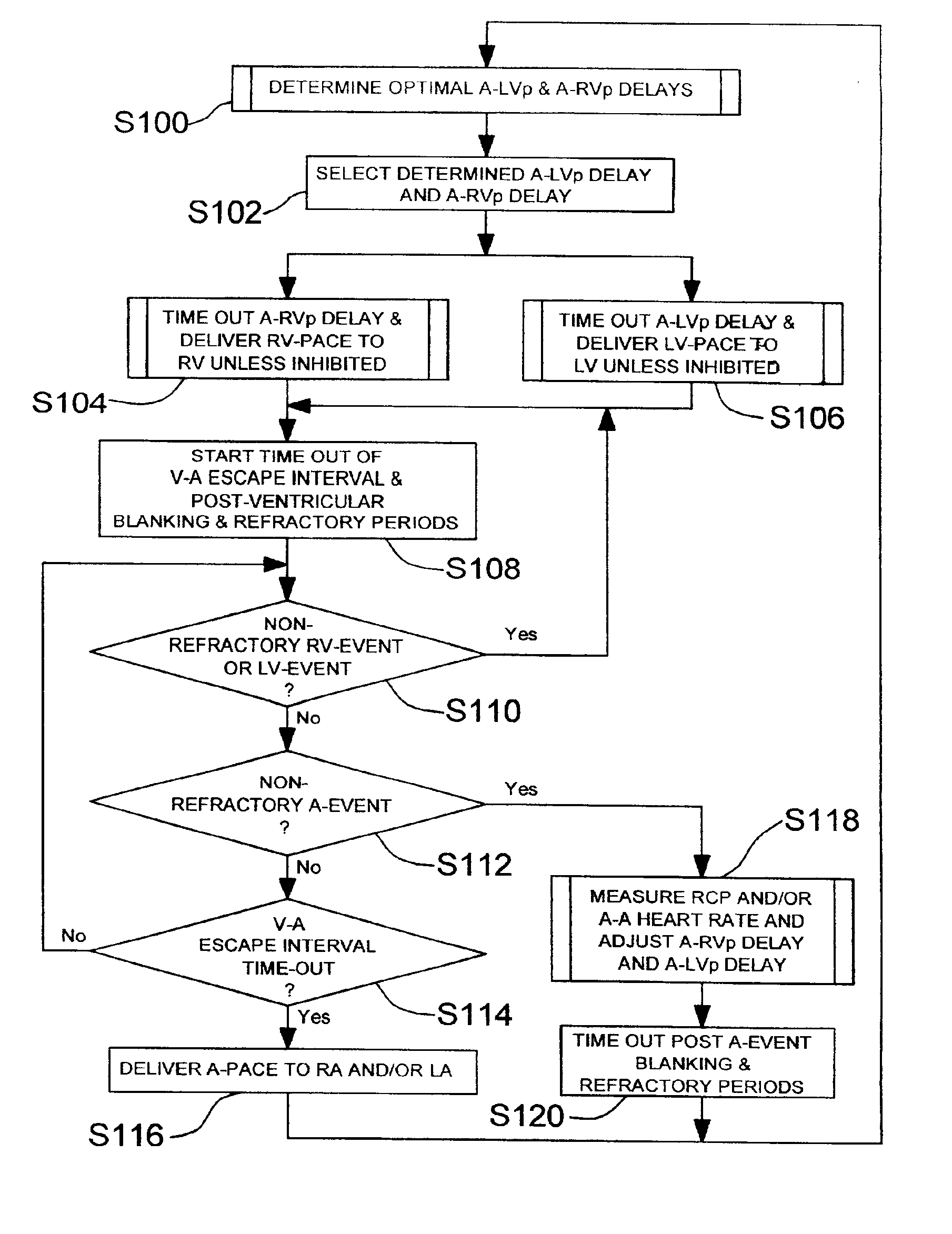
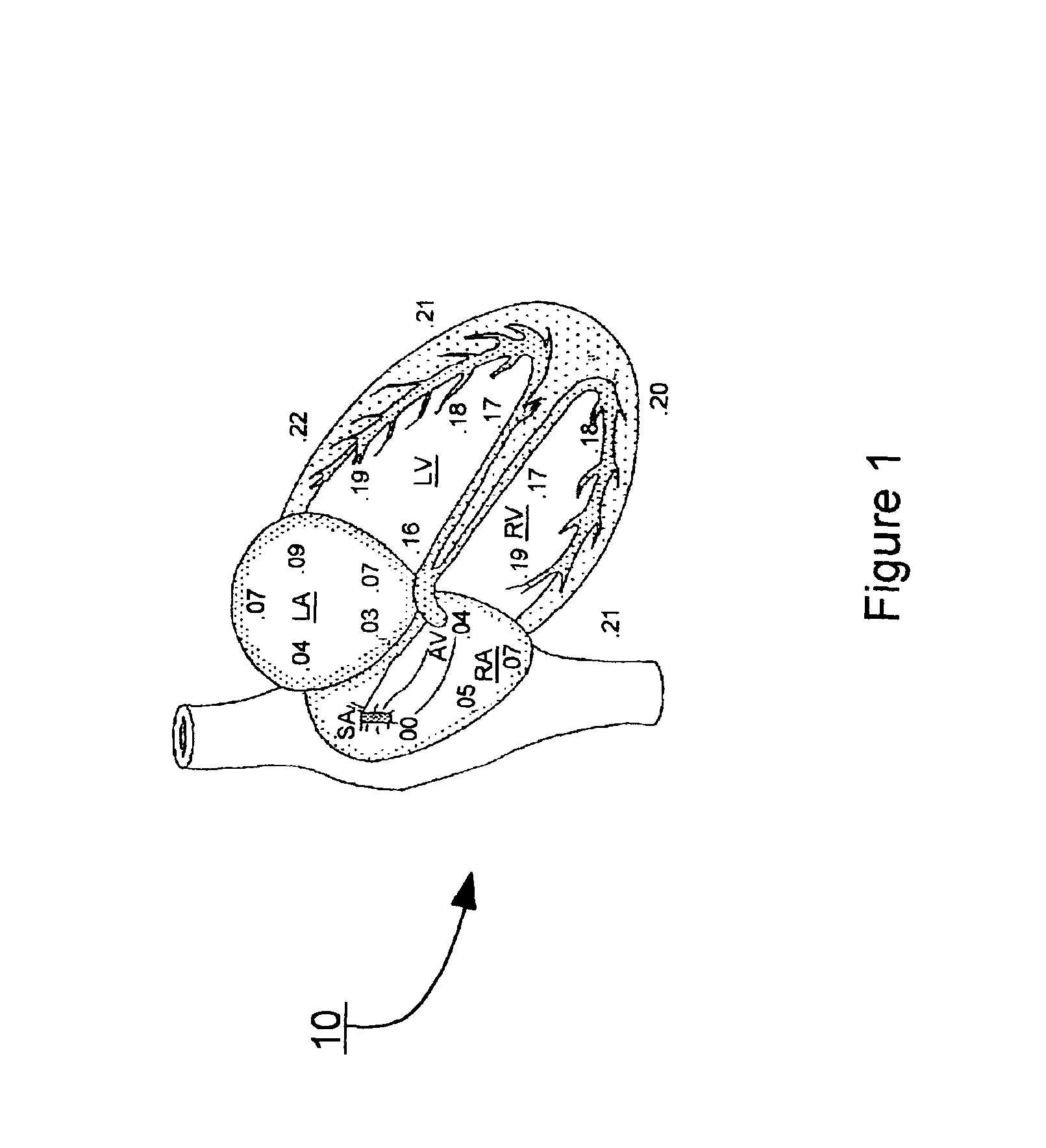
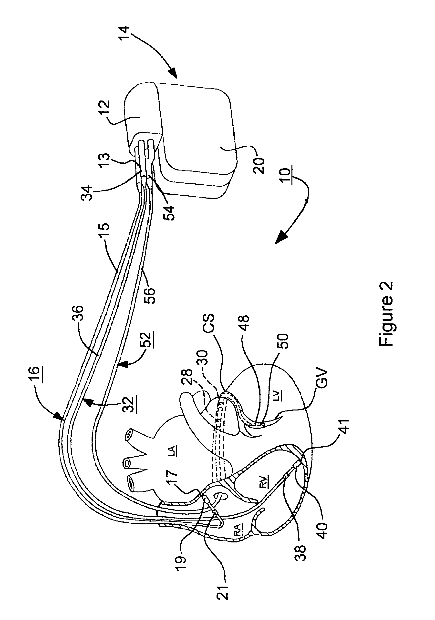
In the following detailed description, references are made to illustrative embodiments for carrying out the invention. It is understood that other embodiments may be utilized without departing from the scope of the invention. For example, the invention is disclosed in detail in FIGS. 2 and 3 in the context of an AV sequential, bi-ventricular, pacing system operating in demand, atrial tracking, and triggered pacing modes in accordance with FIGS. 4 through 7 for restoring synchrony in depolarizations and contraction of the LV and RV in synchronization with atrial paced and / or atrial sensed events. This embodiment of the invention is programmable to operate as a three chamber pacing system having an AV synchronous operating mode for restoring upper and lower heart chamber synchronization and right and left atrial and / or ventricular chamber depolarization synchrony. The system can comprise the capabilities of one of the DDD / DDDR and VDD / DDR bi-ventricular pacing systems but preferably o...


PUM


Abstract

Bi-ventricular cardiac pacing systems and systems for improving cardiac function for heart failure patients that pace and sense in right and left ventricles of the heart and particularly pace in one of the right and left ventricles after an AV delay timed from a preceding atrial event and after a spontaneous depolarization in the other of the right and left ventricles to achieve fusion pacing. An A-RVp delay and an A-LVp delay are each determined from an intrinsic sensed A-RVs delay and an intrinsic A-LVs delay. If the derived A-LVp delay becomes substantially equal to or shorter than the intrinsic A-RVs delay, then the A-RVp delay is decremented to be shorter than the A-LVp delay. Bi-ventricular pacing of the RV and LV is then established closely timed to the intrinsic RV and LV depolarizations.

Description

FIELD OF THE INVENTIONThe present invention pertains to bi-ventricular cardiac pacing systems for improving cardiac function for heart failure patients that pace and sense in right and left ventricles of the heart and particularly pace in one of the right and left ventricles after an AV delay timed from a preceding atrial sense or atrial pace pulse characterized as an A-RVp delay and an A-LVp delay that are each determined from the intrinsic sensed A-RVs delay or the A-LVs delay to effect a fusion of delivered left ventricular pacing pulses with intrinsic depolarizations of the right ventricle.BACKGROUND OF THE INVENTIONDual chamber pacing systems operating in the multi-programmable, VDD, DDD and DDDR pacing modes have been widely adopted in implantable dual chamber pacemakers and certain implantable cardioverter / defibrillators (ICDs) for providing atrial and ventricular synchronized pacing on demand. A DDD pacemaker implantable pulse generator (IPG) includes an atrial sense amplifi...

Claims


Application Information

Patent Timeline
Patent Type & Authority Patents(United States)
IPC IPC(8): A61N1/362A61N1/368A61N1/365
CPCA61N1/3627A61N1/3684A61N1/3682A61N1/36842A61N1/36843
Inventor HILL, MICHAEL R. S.
Owner MEDTRONIC INC
Why Eureka
Try Eureka