
Contrast-agent enhanced color-flow imaging

Inactive Publication Date: 2005-11-29
KONINKLIJKE PHILIPS ELECTRONICS NV
View PDF11 Cites 16 Cited by
  • Summary
  • Abstract
  • Description
  • Claims
  • Application Information

AI Technical Summary

Benefits of technology

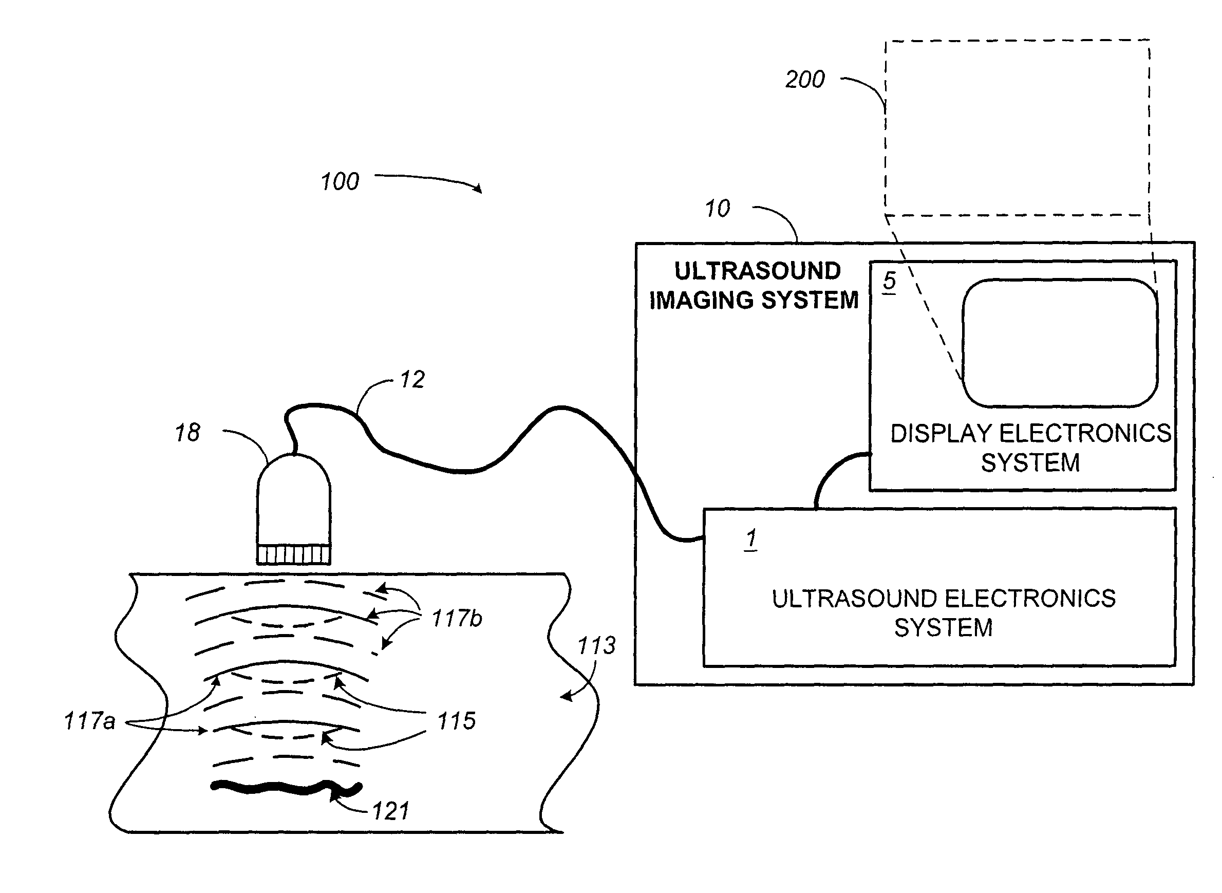
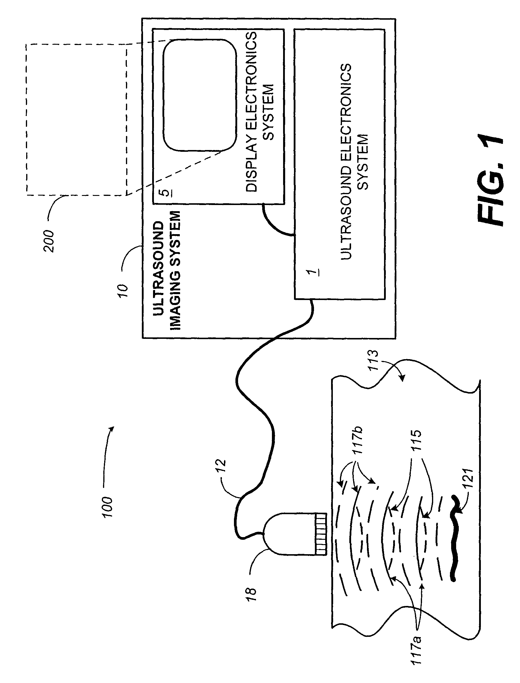
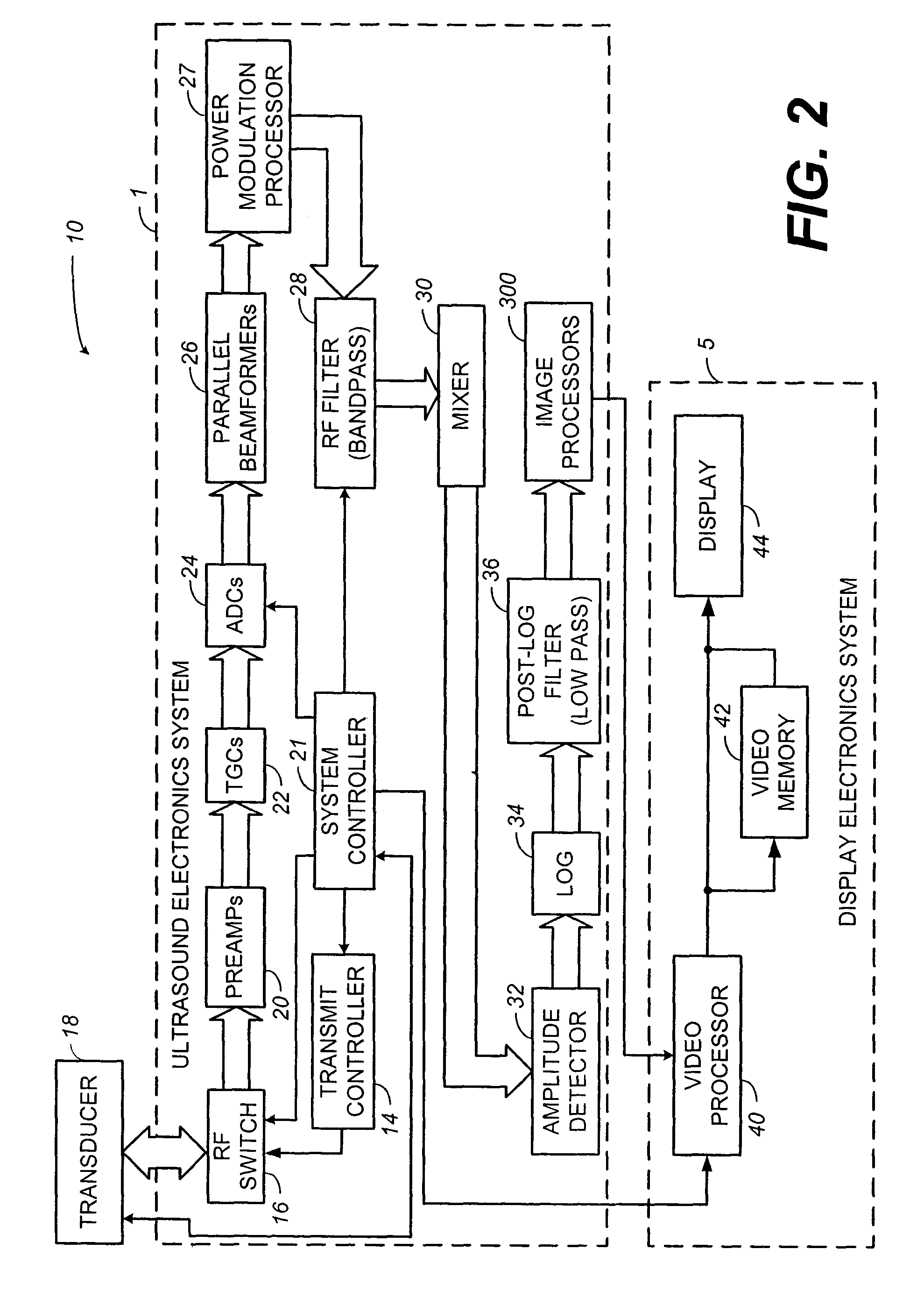
[0012]The present disclosure relates to apparatus and methods for imaging contrast agents within a patient's body. More specifically, the disclosure relates to a system and method that improves contrast-agent enhanced-diagnostic evaluations by applying a color-flow processing algorithm to ultrasonic-response data generated and processed by a power-modulation technique selected for the ability to suppress tissue signals. The method generally comprises introducing one or more contrast agents into the body, transmitting power-modulated ultrasonic pulses into the body, receiving echoed signals from the body, processing the received data (in the received echo signals) to suppress tissue-response signals, processing the received contrast-agent signals with a color-flow algorithm, examining the color-flow processed contrast-agent signals over time to generate a contrast-agent velocity estimate, and generating a color-encoded display of contrast-agent velocities.
[0013]In one embodiment, the ultrasound signal comprises a plurality of signal lines that have been modulated to have different transmit characteristics. The step of processing the received data to suppress tissue-motion generated response signals comprises repetitively applying a weighted finite-impulse-response (FIR) filter to slow-time samples to substantially remove tissue signals and applying the received sequence to a one-zero clutter filter. This has the effect of removing signals from stationary contrast-agent bubbles, while passing signals generated by moving contrast-agent bubbles.

Problems solved by technology

Current color-flow imaging techniques have disadvantages in that it is difficult to obtain diagnostic quality images from patients that have a poor acoustic window (i.e., patients having a relatively large volume of tissue between their skin and their rib cage for heart related studies).
In addition, it is often difficult to separate desired blood-velocity signals from artifacts that result from moving tissue.
This problem is most severe when there is not a relatively large difference in velocity between the tissue and the blood contained therein.
However, single-pulse excitation techniques together with harmonic imaging suffer from poor microbubble-to-tissue ratios as large tissue generated integer-harmonic signals mask the signals generated by the contrast agent.
Although the above-described techniques work well in removing the influence of stationary tissue, flash artifacts from moving tissue can degrade the resultant images.
In particular, this movement causes decorrelation of the received echoes that is not compensated by typical processing techniques.
This degradation can be substantial, particularly where the heart is being imaged due to its frequent and rapid motions.
However, this technique assumes perfectly linear tissue movement and therefore is not completely effective in removing the moving tissue signals.

Method used


Image

  • Contrast-agent enhanced color-flow imaging
  • Contrast-agent enhanced color-flow imaging
  • Contrast-agent enhanced color-flow imaging

Examples


Embodiment Construction

[0028]The present disclosure generally relates to contrast imaging. According to one aspect of the invention, a contrast-agent detection technique is used together with a tissue-signal suppression technique to image contrast-agent concentrations within blood vessels of contrast-agent perfused tissue. In another aspect of the invention, a tissue-motion velocity signal is isolated and used to correct blood-flow velocity information relative to the tissue rather than relative to the transducer. In either case, a color-flow processor is used together with a clutter filter to generate a signal representing contrast-agent velocities. The combination of the tissue-suppression feature of power modulation with flow-estimation feature of color-flow processing makes it possible to differentiate relatively slow-moving blood from moving tissue. Some exemplar clinical applications may include coronary-artery imaging, coronary-flow reserve assessment, blood-perfusion imaging, and tumor detection b...


PUM


Abstract

Systems and methods for enhanced color-flow imaging of contrast-agent perfused blood vessels and other tissues within a patient's body are disclosed. The method generally comprises, introducing one or more contrast agents into the body, power-modulating transmit pulses into the body, receiving echoes from the body, processing the received echoes to reduce tissue-generated echoes and echoes from stationary contrast agent, using a color-flow processor to generate a color-encoded display responsive to contrast-agent motion. The method may be implemented by a system with a an excitation-signal source, a transducer, an ultrasound-processing system having multiple image processors including a color-flow processor, as well as, a clutter filter, and an arbiter, and a display-processing system.

Description

CROSS REFERENCE TO RELATED APPLICATION[0001]This is a continuation of prior application Ser. No. 10 / 134,164, filed Apr. 26, 2002, now U.S. Pat. No. 6,638,228.FIELD OF THE INVENTION[0002]The present disclosure relates to ultrasonic imaging. More particularly, a system and method for improved contrast-agent enhanced-diagnostic evaluations are disclosed.BACKGROUND OF THE INVENTION[0003]Ultrasonic imaging has quickly replaced conventional X-rays in many clinical applications because of its image quality, safety, and low cost. Ultrasonic images are typically formed through the use of phased or linear-array transducers which are capable of transmitting and receiving pressure waves directed into a medium such as the human body. These ultrasonic transducers may be further assembled into a housing, which may contain control electronics, the combination of which forms an ultrasonic probe. Ultrasonic probes are used along with an ultrasonic transceiver to transmit and receive pressure waves th...

Claims


Application Information

Patent Timeline
IPC IPC(8): A61B8/00A61B8/06G01S7/52G01S15/10G01S15/89
CPCA61B8/06A61B8/13A61B8/481G01S7/52038G01S7/52039G01S15/102G01S15/8981
Inventor BROCK-FISHER, GEORGE A.PERRY, JODI L.T.RAFTER, PATRICK G.POLAND, MCKEE DUNN
Owner KONINKLIJKE PHILIPS ELECTRONICS NV
Learn More