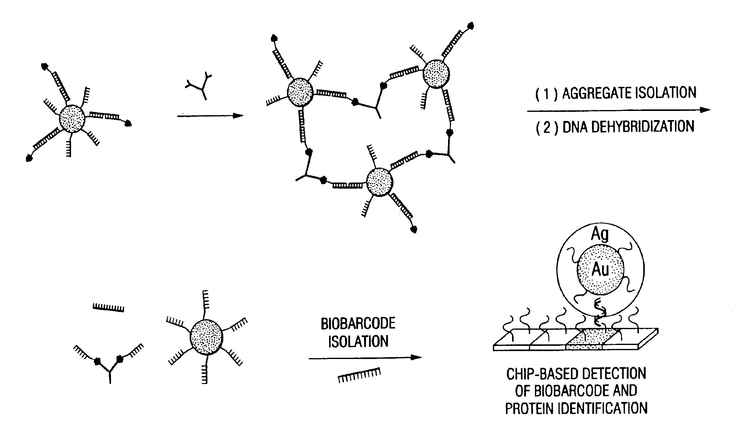
Bio-barcodes based on oligonucleotide-modified nanoparticles
a technology of oligonucleotide and nanoparticles, applied in the field of biobarcodes based on oligonucleotidemodified nanoparticles, can solve the problems of time-consuming, difficult, expensive, etc., and achieve the effect of detecting multiple simultaneously
- Summary
- Abstract
- Description
- Claims
- Application Information
AI Technical Summary
Problems solved by technology
Method used
Image
Examples
example 1
Preparation of Oligonucleotide-Modified Gold Nanoparticles
A. Preparation of Gold Nanoparticles
[0154]Oligonucleotide-modified 13 nm Au particles were prepared by literature methods (˜110 oligonucleotides / particle)18-20. Gold colloids (13 nm diameter) were prepared by reduction of HAuCl4 with citrate as described in Frens, Nature Phys. Sci., 241, 20 (1973) and Grabar, Anal. Chem., 67, 735 (1995). Briefly, all glassware was cleaned in aqua regia (3 parts HCl, 1 part HNO3), rinsed with Nanopure H2O, then oven dried prior to use. HAuCl4 and sodium citrate were purchased from Aldrich Chemical Company. An aqueous solution of HAuCl4 (1 mM, 500 mL) was brought to a reflux while stirring, and then 50 mL of a 38.8 mM trisodium citrate solution was added quickly, which resulted in a change in solution color from pale yellow to deep red. After the color change, the solution was refluxed for an additional fifteen minutes, allowed to cool to room temperature, and subsequently filtered through a Mi...
example 2
Preparation of Hapten-Modified Oligonucleotides
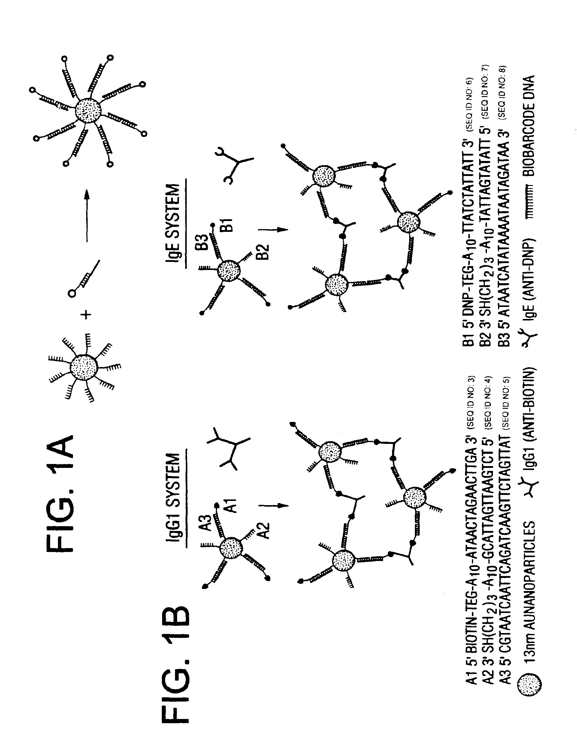
[0160]Hapten-modified oligonucleotides were prepared with a biotin-triethylene glycol phosphoramidite for A1 and 2,4-dinitrophenyl-triethylene glycol phosphoramidite for B1 (Glen research) using standard solid-phase DNA synthesis procedures.21
[0161]Biotin modified oligonucleotides were prepared using the following protocol: A CPG-bound, detritylated oligonucleotide was synthesized on an automated DNA synthesizer (Expedite) using standard procedures21. The CPG-cartridge was then removed and disposable syringes were attached to the ends. 200 μL of a solution containing 20 μmole of biotin-triethylene glycol phosphoramidite in dry acetonitrile was then mixed with 200 μL of standard “tetrazole activator solution” and, via one of the syringes, introduced into the cartridge containing the oligonucleotide-CPG. The solution then was slowly pumped back and forth through the cartridge for 10 minutes and then ejected followed by washing with dry a...
example 3
Assay Using Nanoparticle Complex Probes
[0162]The Oligonucleotide-modified 13 nm gold particles were prepared as described in Example 1. Hapten-modified oligonucleotides were prepared as described in Example 2 with a biotin-triethylene glycol phosphoramidite for A1 and 2, 4-dinitrophenyl-triethylene glycol phosphoramidite for B1 (Glen research) using standard solid-phase DNA synthesis procedures.21 The PBS buffer solution used in this research consists of 0.3 M NaCl and 10 mM phosphate buffer (pH 7). IgE and IgG1 were purchased from Sigma Aldrich (Milwaukee, Wis.) and dissolved in 0.3 M PBS buffer with 0.05% Tween 20 (final concentration: 4.3×10⁻⁸ b / μl) and background proteins (10 ug / ml of lysozyme, 1% bovine serum albumin, and 5.3 ug / ml of anti-digoxin; 10 uL of each) prior to use.
[0163]To prepare the probes, the oligonucleotide modified particles (13 nM, 300 μL) were hybridized with hapten-modified complementary oligonucleotides (10 μL of 10 μM) and biobarcode DNA (10 μL of 10 μM) ...
PUM
| Property | Measurement | Unit |
|---|---|---|
| diameter | aaaaa | aaaaa |
| total volume | aaaaa | aaaaa |
| temperature | aaaaa | aaaaa |
Abstract
Description
Claims
Application Information
 - R&D
- Intellectual Property
- Life Sciences
- Materials
- Tech Scout
- Unparalleled Data Quality
- Higher Quality Content
- 60% Fewer Hallucinations
Browse by: Latest US Patents, China's latest patents, Technical Efficacy Thesaurus, Application Domain, Technology Topic, Popular Technical Reports.
© 2025 PatSnap. All rights reserved.Legal|Privacy policy|Modern Slavery Act Transparency Statement|Sitemap|About US| Contact US: help@patsnap.com


