
Black blood angiography method and apparatus

a black blood angiography and black blood technology, applied in the field of medical imaging arts, can solve the problems of complicated blood vessel segmentation of bba images, inaccurate wba lumen imaging, and degrade the quality of life, so as to facilitate better visualization of the vascular system, accurate vessel lumen information, and remove non-vascular contrast

Inactive Publication Date: 2006-03-28
KONINKLIJKE PHILIPS ELECTRONICS NV +1
View PDF17 Cites 159 Cited by
  • Summary
  • Abstract
  • Description
  • Claims
  • Application Information

AI Technical Summary

Benefits of technology

[0014]One advantage of the present invention is that it enables automated vessel segmentation of black blood angiographic images.
[0015]Another advantage of the present invention is that it removes interfering non-vascular structures having intensities that are similar to the black blood intensity from the black blood image.
[0016]Yet another advantage of the present invention is that it substantially removes non-vascular contrast from a black blood angiographic image to facilitate better visualization of the vascular system using three dimensional, two dimensional, and projection image representations.
[0017]Still yet another advantage of the present invention is that it advantageously retains the sharp vascular edges and accurate vessel lumen information typically achieved by black blood data acquisition during the vessel segmentation processing.
[0018]Numerous additional advantages and benefits of the present invention will become apparent to those of ordinary skill in the art upon reading the following detailed description of the preferred embodiment.

Problems solved by technology

Catastrophic medical events such as heart attacks and strokes that result from underlying vascular problems are a leading cause of death in the United States.
Many Americans also suffer from chronic vascular diseases which degrade quality of life.
Inaccurate WBA lumen imaging is particularly problematic from a clinical perspective because many vascular diseases involve blood vessel stenosis resulting from coating or other degradation of the inside vessel walls.
However, BBA suffers from several disadvantages relating to the post-acquisition image processing.
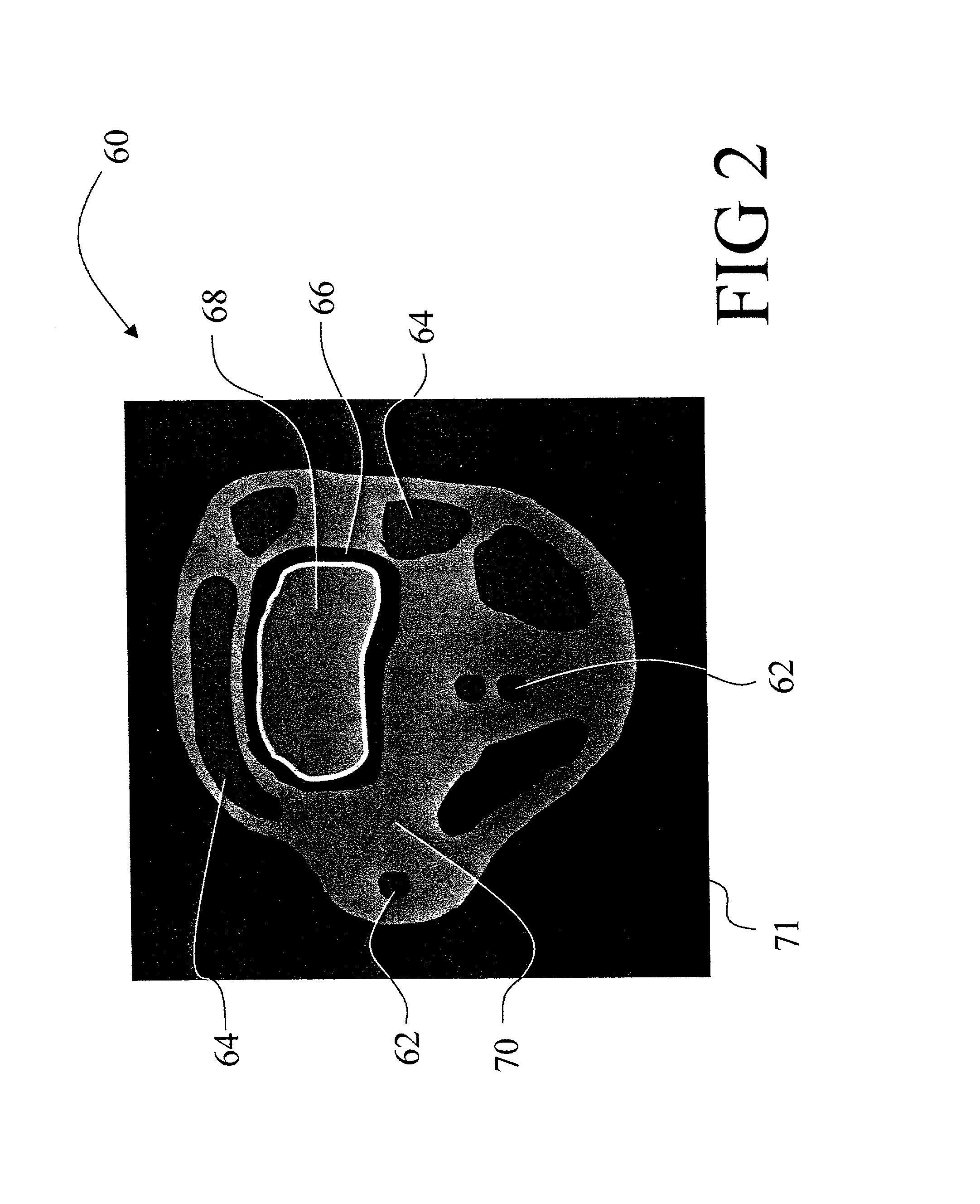
Blood vessel segmentation of BBA images is complicated because the arterial and venous vascular systems are imaged simultaneously and because, in addition to the vascular structures, other non-vascular regions such as air pockets and bone surfaces typically also appear with dark contrast in the BBA images.
Past vessel segmentation methods have proven unsatisfactory for overcoming the problems and generating unambiguous angiographic images from acquired BBA data.

Method used


Image

  • Black blood angiography method and apparatus
  • Black blood angiography method and apparatus
  • Black blood angiography method and apparatus

Examples


Embodiment Construction

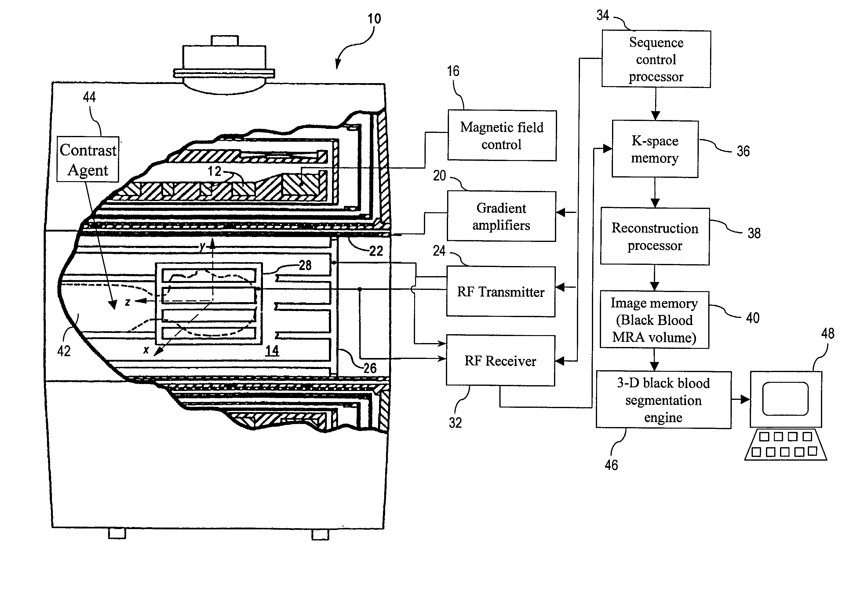
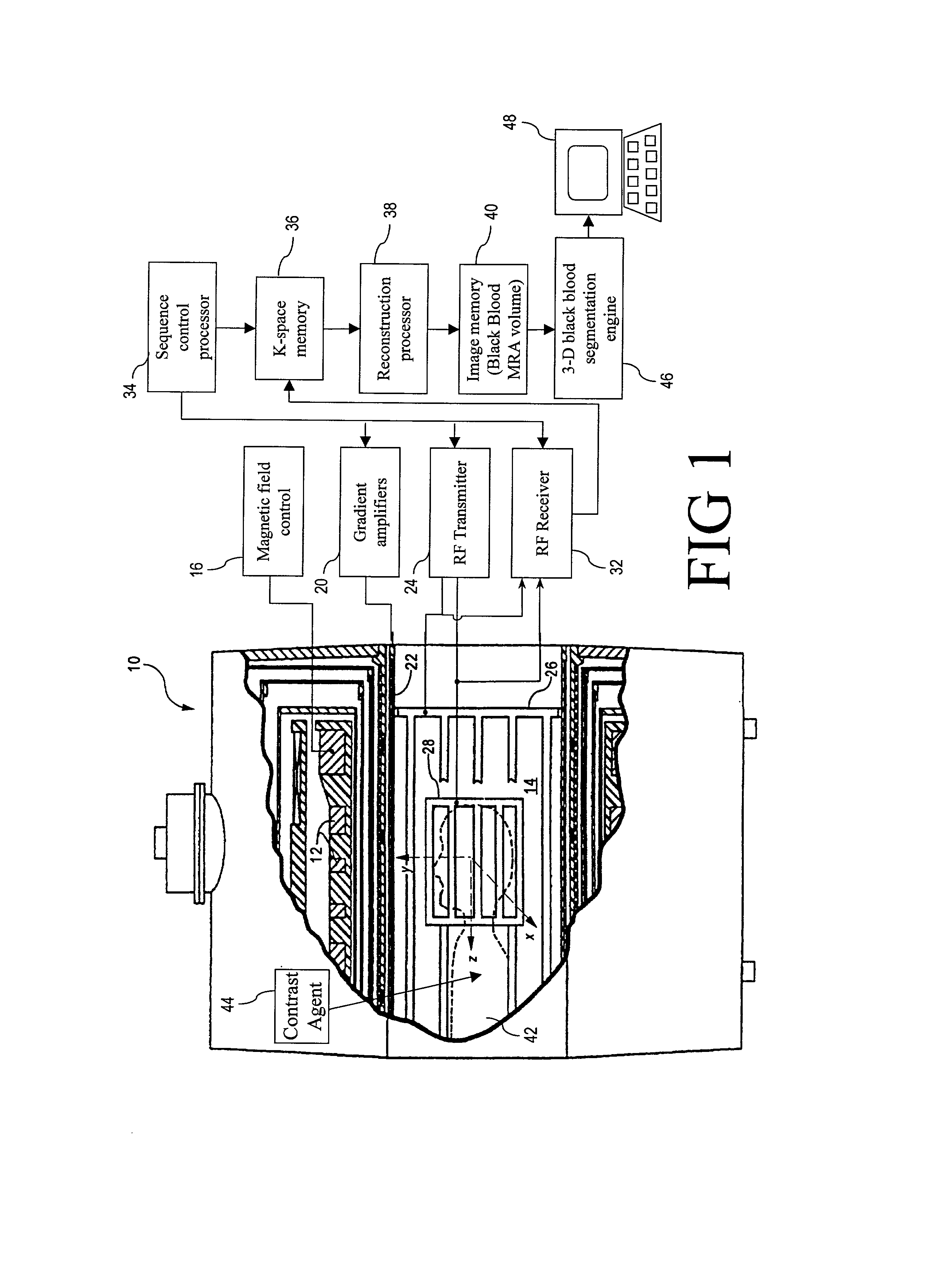
[0060]With reference to FIG. 1, a magnetic resonance imaging system that suitably practices black blood angiographic (BBA) imaging in accordance with an embodiment of the invention is described. Although the invention is described herein with respect to a magnetic resonance imaging embodiment, those skilled in the art will appreciate that the invention is applicable to a broad range of angiographic modalities and techniques in which the angiographic image is of the black blood type. These modalities include but are not limited to contrast-enhanced magnetic resonance angiography, non-contrast enhanced magnetic resonance angiography, and computed tomographic angiography.

[0061]With reference to FIG. 1, a magnetic resonance imaging (MRI) scanner 10 typically includes superconducting or resistive magnets 12 that create a substantially uniform, temporally constant main magnetic field B0 along a z-axis through an examination region 14. Although a bore-type magnet is illustrated in FIG. 1, ...


PUM


Abstract

To produce a black body angiographic image representation of a subject (42), an imaging scanner (10) acquires imaging data that includes black blood vascular contrast. A reconstruction processor (38) reconstructs a gray scale image representation (100) from the imaging data. A post-acquisition processor (46) transforms (130) the image representation (100) into a pre-processed image representation (132). The processor (46) assigns (134) each image element into one of a plurality of classes including a black class corresponding to imaged vascular structures, bone, and air, and a gray class corresponding to other non-vascular tissues. The processor (46) averages (320) intensities of the image elements of the gray class to obtain a mean gray intensity value (322), and replaces (328) the values of image elements comprising non-vascular structures of the black class with the mean gray intensity value.

Description

BACKGROUND OF THE INVENTION[0001]The present invention relates to the medical imaging arts. It particularly relates to black-blood angiography (BBA) using the magnetic resonance imaging (MRI) and computed tomography (CT) medical imaging techniques, and will be described with particular reference thereto. However, the invention will also find application in conjunction with other three-dimensional imaging modalities as well as in other imaging arts in which structures or networks with well-defined characteristics are advantageously differentiated from extraneous imaged structures and background noise.[0002]Catastrophic medical events such as heart attacks and strokes that result from underlying vascular problems are a leading cause of death in the United States. Many Americans also suffer from chronic vascular diseases which degrade quality of life.[0003]Angiography relates to the imaging of blood vessels and blood vessel systems, and as such is a powerful medical diagnostic for iden...

Claims


Application Information

Patent Timeline
IPC IPC(8): G06K9/00G01R33/28A61B5/055G01R33/54G01R33/56G01R33/563G06T5/00G06V10/52
CPCG06K9/527G01R33/563G01R33/5608G06V10/52
Inventor SURI, JASJIT S.LIU, KECHENG
Owner KONINKLIJKE PHILIPS ELECTRONICS NV
Learn More