Tissue interface device

a technology of tissue interface and interface device, which is applied in the field of tissue interface device, can solve the problems of obviating the significant disadvantage of prior art devices and methods, and achieve the effects of reducing the number of devices, and improving the quality of tissue interfa

Inactive Publication Date: 2006-05-09
ALTEA THERAPEUTIC CORP +1
View PDF10 Cites 243 Cited by
  • Summary
  • Abstract
  • Description
  • Claims
  • Application Information

AI Technical Summary

Benefits of technology

[0009]The present invention involves positioning the sensor ex vivo, proximate the surface of the organism, and coupling the sensor to the organism via the fluid conducting tapered orifice of the housing. Consequently, oxygen (if necessary) to support the sensor reaction is readily available, allowing for a simpler basic array design, higher signal-to-noise ration, faster response, better linear tracking of the physiological changes in an analyte of interest, and longer life of the sensor. By keeping all of the foreign material of the tissue interface device outside of the body, the auto-immune derived encapsulation and rejection responses naturally occurring with any implanted device never begin.
[0010]Further, by avoiding actual penetration of the body to insert a sensor, needle, or cannula, a significant disadvantage of the prior art devices and methods is obviated by the tissue interface device of the present invention. Also, risks of infection that are present in the prior art systems are dramatically reduced in connection with the present invention because neither sensor implantation is involved nor a membrane breaching connection to a needle or cannula.
[0011]Still further, the tissue interface device of the present invention provides for high interstitial fluid flux rates without causing increased erythema that may prolong skin healing. Additionally, by minimizing the dead volume between the artificial opening in the membrane and the sensor, and hence minimizing the lag time, a further significant disadvantage of the prior art systems and method is obviated by the device of the present invention.

Problems solved by technology

Further, by avoiding actual penetration of the body to insert a sensor, needle, or cannula, a significant disadvantage of the prior art devices and methods is obviated by the tissue interface device of the present invention.

Method used


Image

  • Tissue interface device
  • Tissue interface device
  • Tissue interface device

Examples


Embodiment Construction

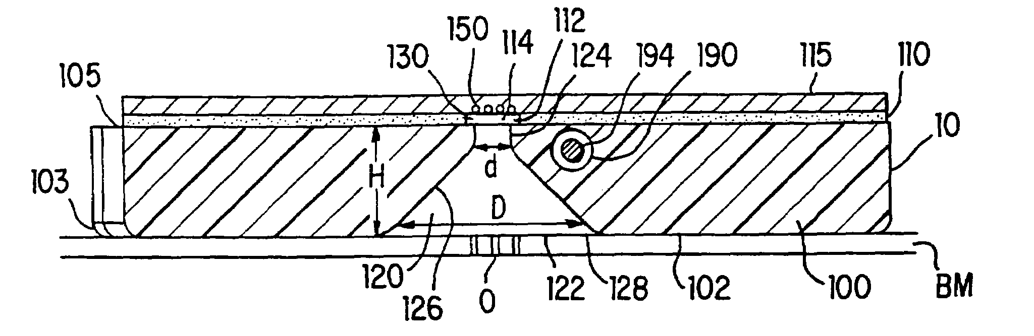
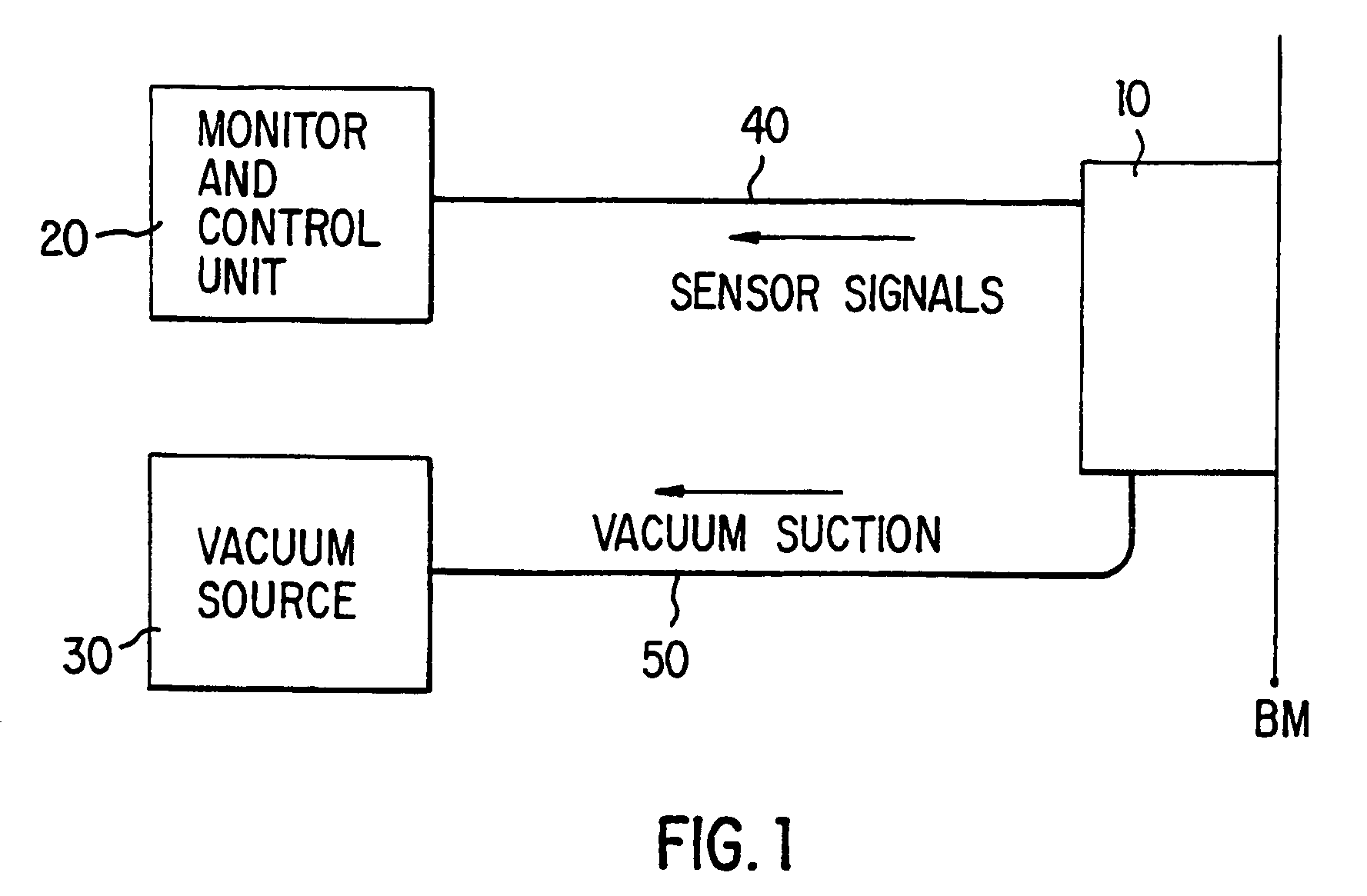
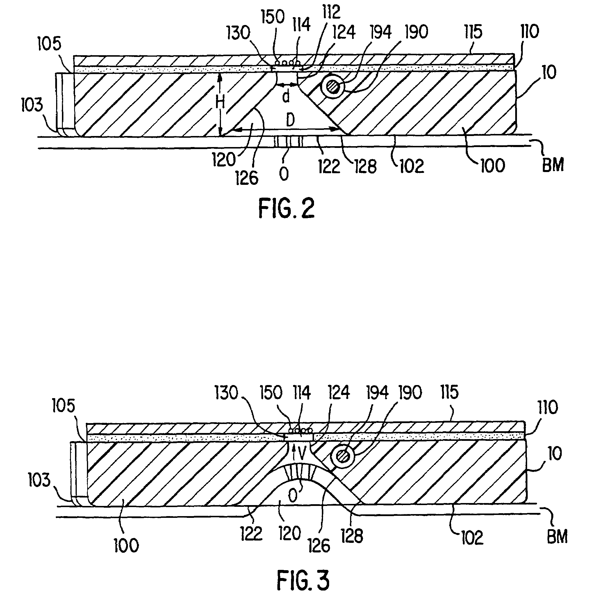
[0022]The present invention may be understood more readily by reference to the following figures and their previous and following description, in which like numbers indicate like parts throughout the figures. It is to be understood that this invention is not limited to the specific devices described, as specific device components as such may, of course, vary. It is also understood that the terminology used herein is for the purpose of describing particular embodiments only and is not intended to be limiting.

[0023]As used in this specification and the appended claims, the singular forms “a,”“an” and “the” may mean one or more than one. For example, “a” sensor may mean one sensor or more than one sensor.

[0024]Ranges may be expressed herein as from “about” or “approximately” one particular value and / or to “about” or “approximately” another particular value. When such a range is expressed, another embodiment comprises from the one particular value and / or to the other particular value. S...


PUM

PropertyMeasurementUnit
diameteraaaaaaaaaa
diameteraaaaaaaaaa
diameteraaaaaaaaaa

Abstract

A tissue interface device (10) suitable for positioning on or about one or more artificial openings in a biological membrane of an organism and for coupling to a monitor and control unit and a vacuum source. The tissue interface device (10) comprises a housing (100), a sensor channel (130), and a sensor (150). The housing (100) defines an orifice (120), the orifice (120) having an open inlet port (122) on the bottom end (102) of the housing (100) and a distal end (124) that is in fluid communication with the sensor channel (130). The orifice (120) is in fluid communication with fluid that flows from the artificial opening formed in the biological membranes. The sensor channel (130) is for coupling to, and fluid communication therewith, the vacuum source. The sensor (150) is positioned in the sensor channel (130) in a flow path of the fluid for sensing a characteristic of the fluid as it flows out from the artificial opening. The sensor generates a sensor signal representative thereof.

Description

[0001]This application claims priority to U.S. Provisional Application No. 60 / 166,481 filed Nov. 19, 1999; U.S. Provisional Application No. 60 / 178,148 filed Jan. 26, 2000; and U.S. Provisional Application No. 60 / 244,568 filed Oct. 31, 2000.BACKGROUND OF THE INVENTION[0002]1. Field of the Invention[0003]The present invention relates to tissue interface devices, and more particularly to a tissue interface device wherein fluid is extracted from a biological tissue and placed into contact with a sensor for sensing a characteristic of the fluid flowing from an artificial opening in a biological membrane.[0004]2. Background Art[0005]Monitoring systems that sample and measure characteristics of fluids from a biological tissue, such as on a human, are well known. Many of these systems involve implanting sensors and related devices into the organism (such as under the skin) in order to obtain fluid samples and make measurements of those samples. Implanted sensors, in addition to the pain cau...

Claims


Application Information

Patent Timeline
Patent Type & Authority Patents(United States)
IPC IPC(8): A61B5/00A61B5/04A61B5/05G01N33/487G01N33/48
CPCA61B5/1486
Inventor FAUPEL, MARK L.KUMAR, KRISHNAFARQUHAR, J. DAVIDVREEKE, MARKSMITH, ALANHATCH, MICHAELWOODS, TERESA
Owner ALTEA THERAPEUTIC CORP
Why Eureka
Try Eureka