

Methods of using immunophilin binding drugs to improve integration and survival of neuronal cell transplants
a neuronal cell transplant and immunophilin technology, applied in the field of neurodegenerative diseases using neuronal treatment, can solve the problems that none of these strategies deliver the needed long-term clinical improvement, and achieve the effect of enhancing the growth, survival and integration capabilities of neuronal cells
- Summary
- Abstract
- Description
- Claims
- Application Information
AI Technical Summary
Benefits of technology
Problems solved by technology
Method used
Image

Examples
example 1
Culture of Cells with Immunophilin Binding Drugs in Vitro
[0057]Surgical pathology fetal specimens were collected and processed in accordance with the University of Pittsburgh Human Tissue Committee guidelines. Human telencephalic tissues (18-21 weeks of gestation) were collected and placed in DMEM medium (Life Technologies Inc., Grand Island, N.Y.). The aspiration method of fetal removal did not allow for identification of specific brain structures except the general lobe architecture. Tissue processing followed a modified mouse brain dissociation protocol. Within 1 hour of collection, the brain material was placed in Ca++-and Mg++-free DPBS (Life Technologies), cleaned of meninges, rinsed and minced. Tissue fragments were incubated in 0.05% trypsin-EDTA with additives (Life Technologies) for 5 min at 37° C. and further dissociated. Trypsin inhibitor (10 mg / ml; Sigma, St. Louis, Mo.) was subsequently added, followed by centrifugation for 5 min at 250×g. After filtering through 70 μm...
example 2
In vivo Grafting of Neuronal Cells.
[0068]Second trimester fetal cells grown in suspension at a density of IXI06 cells / mL in 165 cm2 vented flasks (Costar, Cambridge, Mass.) formed aggregates within 48 h. After one week in suspension culture, the human neuroglial aggregates (average diameter of 200 □m) were transplanted into the striatum of 4 wk old adult male SCID mice (Tac:Icr:Ha (ICR)-scidfDF-Taconic Laboratories, Gerrnantown, N.Y.). Human neuroglial aggregate suspension was centrifuged for 5 min at 250×g, resuspended in serum-free culture media and transferred to a GASTIGHT 1705 syringe (Hamilton Company, Reno, Nev.). Mice were anesthetized with Metafane inhalant. Stereotaxic unilateral injections of approximately 20 ?l of the dense brain aggregate suspension were made into the striatum. The coordinates of injection were: 2.5 mm lateral to lambda, 5 mm anterior to lambda, and 3.5 mm ventral to the dural surface. Eight mice per group were sacrificed at 2, 4, 8, 16, 24 and 32 wk po...
example 3
Survival of Second Trimester Fetal Cells in Vitro
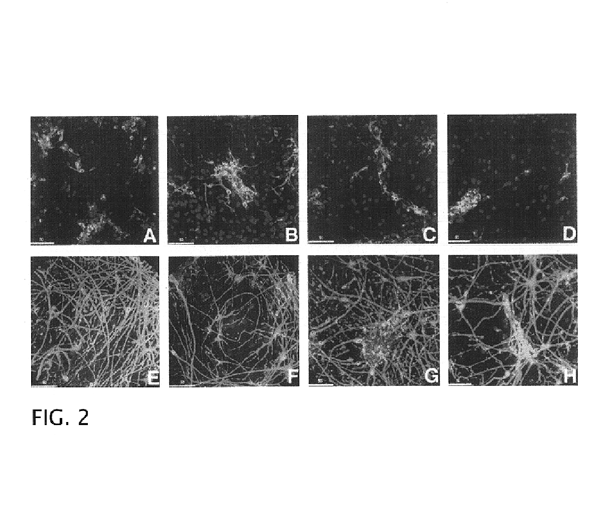
[0073]Single cell suspensions of freshly dissociated second trimester human fetal neuroglia grown at 1×10⁶ cells / nL in serum-free culture media formed spheroid aggregates within 24-48 h (FIG. 6A). By phase-contrast analysis, these aggregates consisted of small, bright cells surrounding a core of one or two larger and darker cells with a morphology consistent with macrophages / microglia By IF analysis, the large cells at the core of the aggregates were confirmed to be of macrophage lineage (e.g. positive for RCA-1) (FIG. 6B). At early time points, within the first two weeks, the aggregates had a diameter between 100 and 200 μm and consisted of approximately 20-30% cells positive for neuronal markers (e.g. PGP 9.5 ), 20% cells positive for astroglial markers (GFAP) and 5% cells positive for microglial markers (e.g. RCA-1).
[0074]During the first two weeks in culture, the aggregates continued to grow in size while their cores became more o...
PUM
| Property | Measurement | Unit |
|---|---|---|
| Density | aaaaa | aaaaa |
| Affinity | aaaaa | aaaaa |
Abstract
Description
Claims
Application Information