Methods of modulating localization and physiological function of IP3 receptors
- Summary
- Abstract
- Description
- Claims
- Application Information
AI Technical Summary
Problems solved by technology
Method used
Image
Examples
example 1
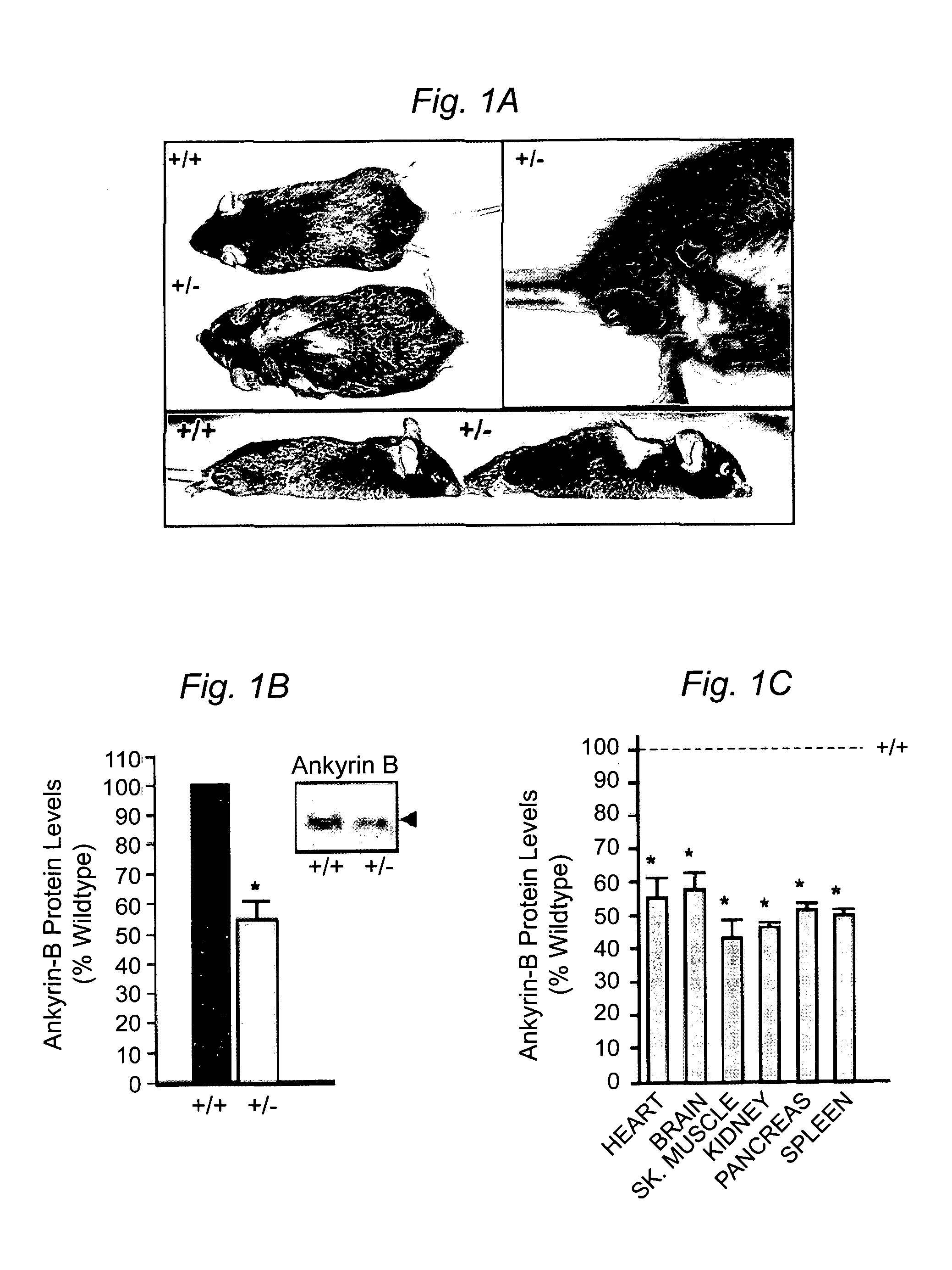
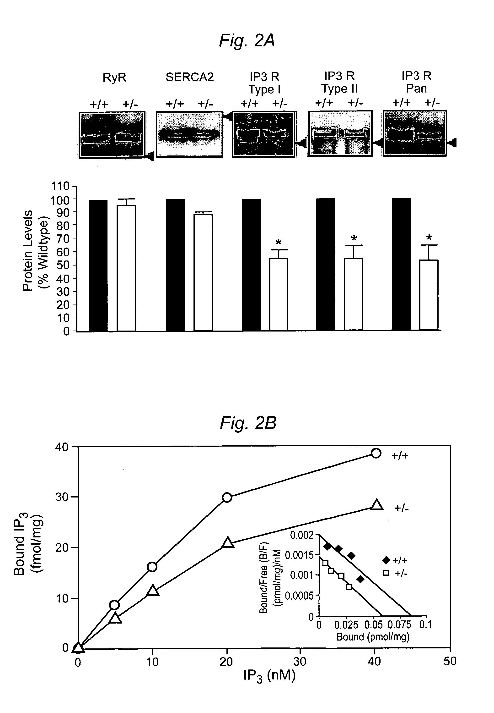
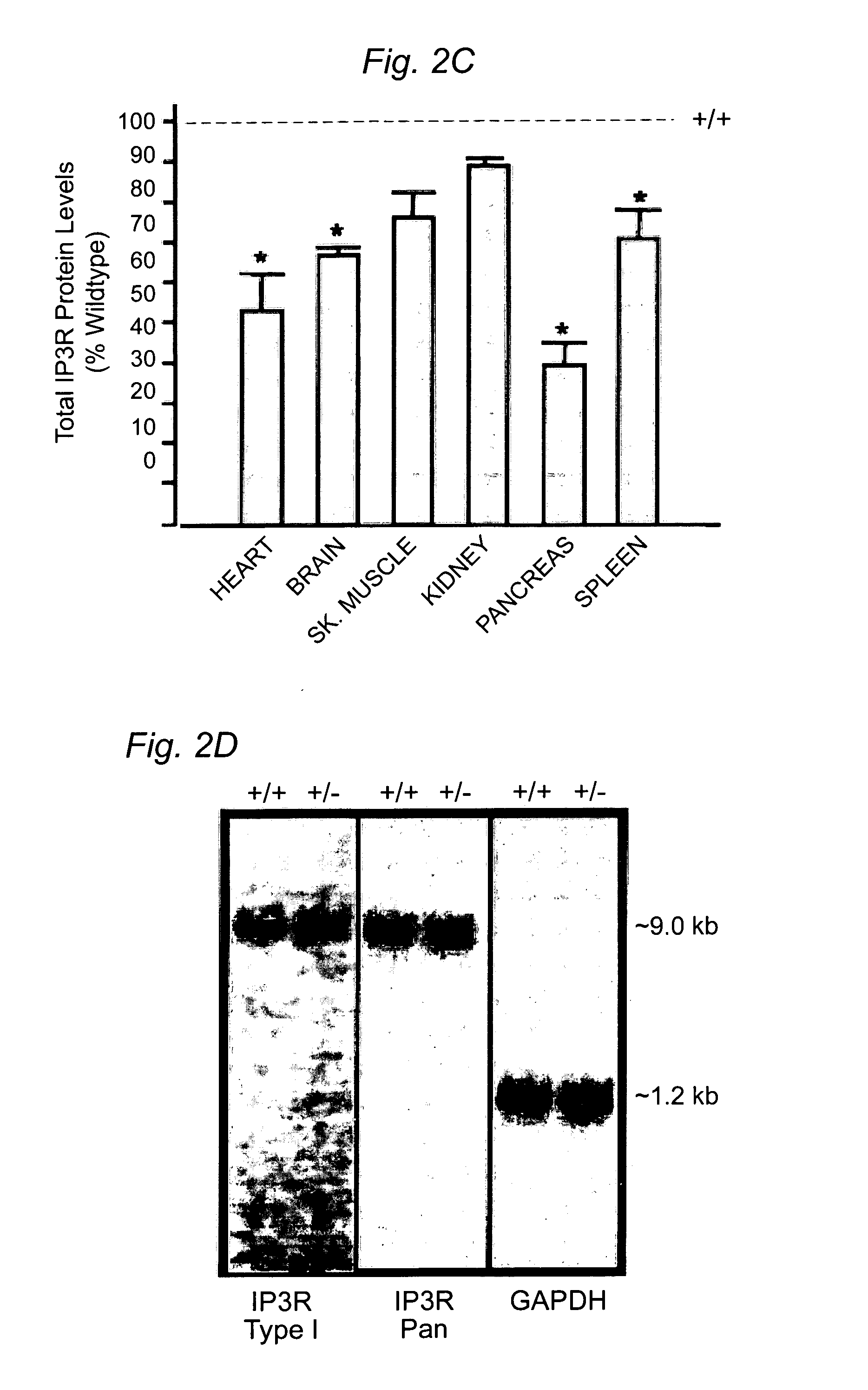
Cardiac Arrhythmia and Abnormal Glucose Regulation in Mice Heterozygous for Null Mutation in Ankyrin-B
Experimental Procedures
[0056]Cell Culture and Calcium Imaging. Neonatal myocytes were isolated and calcium imaging was performed as described (Tuvia et al, J. Cell Biol. 147:995–1008 (1999)). Briefly, calcium imaging in spontaneously contracting 5–6 day old myocytes was performed using fluo-3 / AM (Molecular Probes). Cells were loaded with 10 μM fluo-3 / AM for 30 minutes at 37° C. and washed Images were collected at 8 frames / sec and assembled using Adobe Premiere.
[0057]Immunofluorescence and Immunoblotting. Primary cultures and 8–10 μm tissue sections were analyzed using the following antibodies: insulin, glucagon, α-actinin, DHPR (Sigma), IP3R type I, II, III and a Pan antibody, RyR type II (RyR2), SR / ER Ca2+ATPase (SERCA2; Affinity Bioreagents) or AnkB. For islet area, sections were stained using H & E or a glucagon antibody to visualize the α-cells (Lee and Laychock, Biochem. Pharma...
example 2
Ankyrin-B (+ / −) Mice Display Reduced Response to Phenylephrine and Endothelin-1 on Heart Rate
Ankyrin-B (+ / −) Mice Display Reduced Heart Rate
[0096]To test ankyrin-B heterozygous mice for potential defects in Gαq signaling, ECG radiotransmitter implants were implanted in the abdomen of ankyrin-B (+ / −) mice as well as wildtype littermate controls. These probes allow the recording of real time ECG and thus heart rate recordings in conscious, non-anethestized mice. 24 hour recordings of wildtype mice compared to heterozygote animals show overall bradycardia in the heterozygote (FIG. 9).
Ankyrin-B Mice Display Decreased Sensitivity to Alpha-Adrenergic Stimulation
[0097]Ankyrin-B (+ / −) mice and wildtype littermates were injected with the alpha-adrenergic receptor agonist phenylephrine (PE, an α-adrenergic agonist). As expected, when wildtype mice where intraperitoneally injected with phenylephrine (3 mg / kg) there was a rapid decrease in heart rate, a sustained plateau, followed by a slow ret...
example 3
Description of Ankyrin-B DNA Constructs
[0101]Construct generation. 220 kDa ankyrin-B and 190 kDa ankyrin-G chimeric EGFP expression constructs were engineered using common molecular techniques. Briefly, an internal EcoRI site in ankyrin-B was removed by Quickchange PCR (Stratagene; La Jolla, Calif.). Next, pEGFPC2 and pEGFPN3 were modified to create a novel PmeI site in the pEGFP multiple cloning site (3′). The membrane-binding domain of 220 kDa ankyrin-B and 190 kDa ankyrin-G were amplified by PCR to engineer a 5′ EcoRI site and 3′ AscI site resulting in a three amino acid linker (Gly-Ala-Pro) between the membrane- and spectrin-binding domains. The spectrin-binding domains of 220 kDa ankyrin-B and 190 kDa ankyrin-G, (which lacks the serine / threonine rich insert and tail of 270 kDa ankyrin-G) were amplified by PCR with 5′ AscI and 3′ PacI sites resulting in a three amino acid linker (Leu-Ile-Asn) between and spectrin-binding and death / C-terminal domains. Finally, the death / C-termina...
PUM
| Property | Measurement | Unit |
|---|---|---|
| Atomic weight | aaaaa | aaaaa |
Abstract
Description
Claims
Application Information
 - R&D
- Intellectual Property
- Life Sciences
- Materials
- Tech Scout
- Unparalleled Data Quality
- Higher Quality Content
- 60% Fewer Hallucinations
Browse by: Latest US Patents, China's latest patents, Technical Efficacy Thesaurus, Application Domain, Technology Topic, Popular Technical Reports.
© 2025 PatSnap. All rights reserved.Legal|Privacy policy|Modern Slavery Act Transparency Statement|Sitemap|About US| Contact US: help@patsnap.com


