Limited-angle frequency-distance resolution recovery in nuclear medicine imaging

a frequency-distance resolution and limited-angle technology, applied in the field of diagnostic imaging art, can solve the problems of depth-dependent blurring and uncertainty, non-stationary convolution which is difficult to deconvolve, prior art resolution recovery techniques do not work on partial data sets, etc., and achieve the effect of accurate restoration of limited-angle data sets

Inactive Publication Date: 2007-01-02
MARCONI MEDICAL SYST
View PDF6 Cites 6 Cited by
  • Summary
  • Abstract
  • Description
  • Claims
  • Application Information

AI Technical Summary

Benefits of technology

[0009]One advantage of the present invention is that it accurately restores limited-angle data sets.
[0010]Another advantage of the present invention is that it restores continuously scanned data sets.
[0011]Another advantage of the present invention is that it process

Problems solved by technology

This creates depth-dependent blurring and uncertainty in the resultant image data.
This error is a non-stationary convolution which is difficult to deconvolve.
Although these prior art resolution recovery technique

Method used


Image

  • Limited-angle frequency-distance resolution recovery in nuclear medicine imaging
  • Limited-angle frequency-distance resolution recovery in nuclear medicine imaging
  • Limited-angle frequency-distance resolution recovery in nuclear medicine imaging

Examples


Embodiment Construction

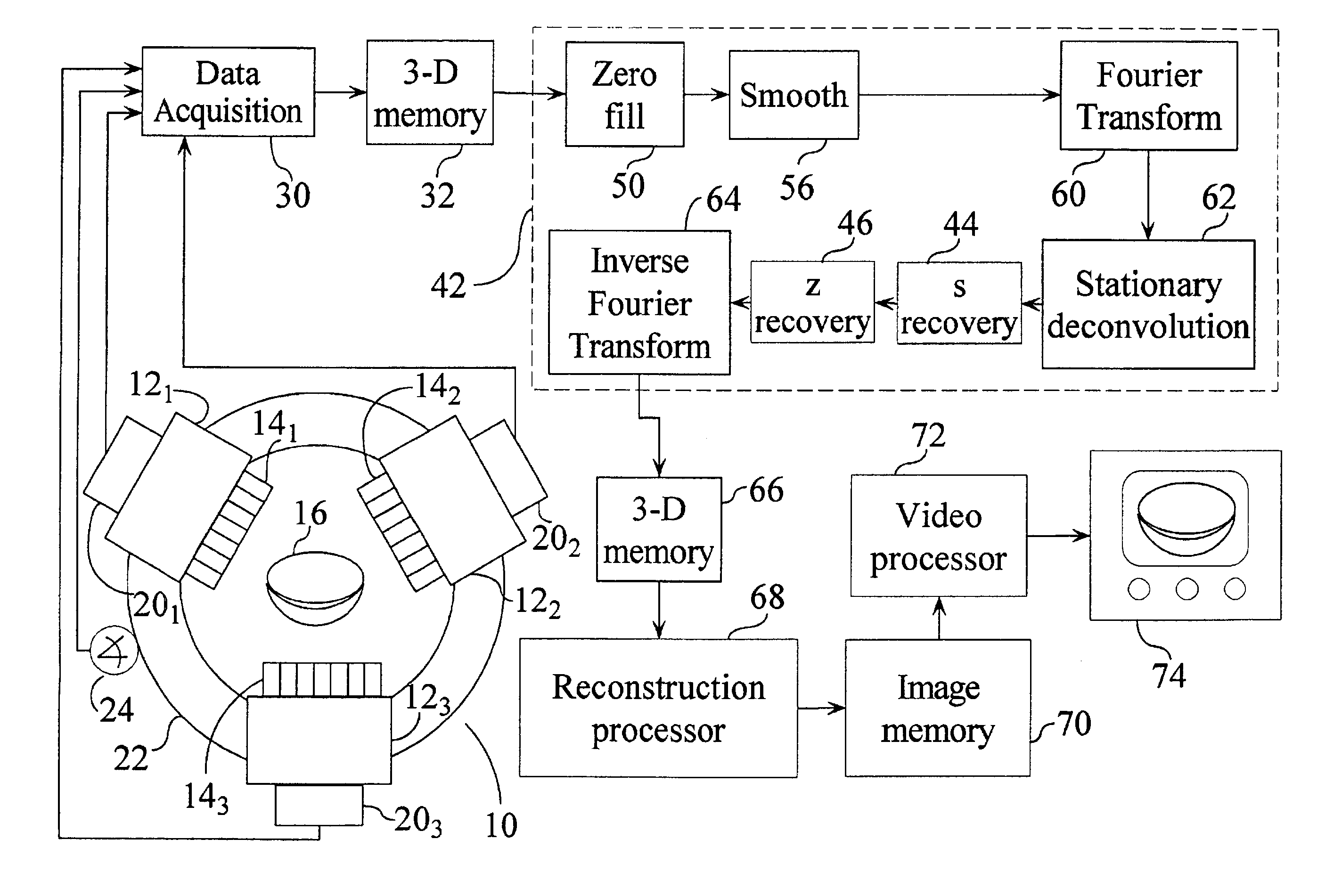
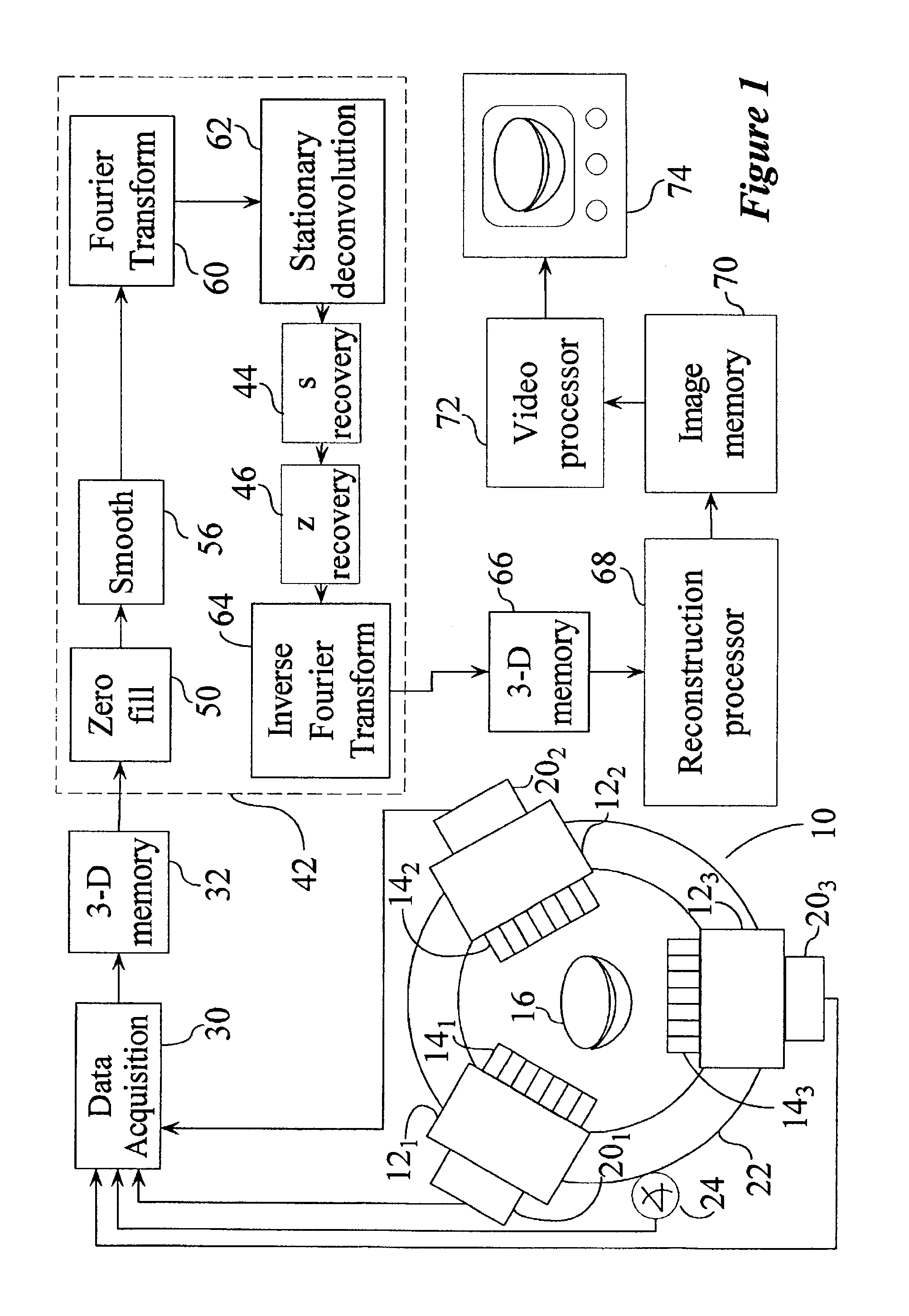
[0017]With reference to FIG. 1, a nuclear camera system 10 includes a plurality of detector heads 12, in the preferred embodiment three detector heads 121, 122, and 123. Two of the three heads are typically used to obtain emission data, while the third head is used to obtain transmission data. Of course, other numbers of detector heads can also be utilized. Each of the detector heads includes a collimator 141, 142, and 143. In the preferred embodiment, the collimators collimate incoming radiation from a subject 16 to parallel rays. However, because the collimators have finite size, each collimator permits rays which lie along a corresponding cone to pass to the detector head. The cone expands with depth into the patient from the detector head.

[0018]Each of the detector heads includes a reconstruction system 201, 202, and 203 which determines the coordinates on a face of the detector head in the longitudinal or z-direction of the patient and the transverse direction across the detect...


PUM


Abstract

A nuclear camera (10) includes a plurality of detector heads (12) which have collimators (14) for fixing the trajectory along which radiation is receivable. A rotating gantry (22) rotates the detector heads around the subject collecting less than 360° of data, e.g., 204° of data. A zero-filling processor (50) generates zero-filled projection views such that the actually collected projection views and the zero-filled projection views span 360°. A smoothing processor (56) smooths an interface between the zero-filled and actually collected projection views. The zero-filled and smoothed views are Fourier transformed (60) into frequency space, filtered with a stationary deconvolution function (62), and Fourier transformed (64) back into real space. The resolution recovered projection data sets in real space are reconstructed by a reconstruction processor (68) into a three-dimensional image representation for storage in an image memory (70).

Description

BACKGROUND OF THE INVENTION[0001]The present invention relates to the diagnostic imaging art. It finds particular application in conjunction with nuclear single photon emission computed tomography (SPECT) medical imaging and will be described with particular reference thereto. However, the invention will also find application in conjunction with other types of non-invasive diagnostic imaging.[0002]Heretofore, diagnostic images have been generated from single and multiple-head nuclear cameras. Typically, a patient positioned in an examination region is injected with a radio pharmaceutical. Heads of the nuclear camera are positioned closely adjacent to the patient to monitor the radio pharmaceutical. Typically, the heads are stepped in increments of a few degrees around the patient until 360° of data have been acquired. That is, projection data along directions spanning 360° are collected. With multiple-head systems, projections along each direction need only be collected with one of ...

Claims


Application Information

Patent Timeline
IPC IPC(8): A61B6/00G01T1/161G06T1/00G06T11/00
CPCG06T11/005G06T2211/436
Inventor HAWKINS, WILLIAM G.
Owner MARCONI MEDICAL SYST
Why Eureka
Try Eureka