Biopsy system having a single use loading unit operable with a trocar driver, a knife driver and firing module

a biopsy system and loading unit technology, applied in the field of single insertion, multiple sample percutaneous biopsy system and method of use, can solve the problems of difficult to determine if pre-palpable breast abnormalities are malignant, difficult to perform mammography, tedious and time-consuming practice, etc., and achieve the effect of reducing the drag on the system

Inactive Publication Date: 2007-03-13
TYCO HEALTHCARE GRP LP
View PDF298 Cites 1328 Cited by
  • Summary
  • Abstract
  • Description
  • Claims
  • Application Information

AI Technical Summary

Benefits of technology

[0014]In accordance with the present disclosure, a biopsy system is provided which includes a single use loading unit (SULU), a knife driver and a trocar driver. The SULU includes a trocar assembly having a tubular trocar defining a tissue receiving basket, a trocar tip having a basket insert fastened to the trocar, a trocar flange secured to the proximal end of the trocar, and an engagement mechanism for connecting the trocar driver to the trocar. The engagement mechanism includes a connecting member in the form of a notched flag. The connecting member is secured to one end of a drive shaft which extends between the trocar driver and the SULU. The notched flag is positioned to engage the trocar flange such that axial movement of the drive shaft effected by the trocar driver is translated to axial movement of the trocar. The connecting member is pivotable out of engagement with the trocar flange to disengage the trocar driver from the trocar to reduce drag on the system during firing of the trocar and enable advancement of the trocar into a target tissue mass at a higher velocity.

Problems solved by technology

The type of biopsy utilized depends in large part on the circumstances present with respect to the patient and no single procedure is ideal for all cases.
However, definitive tissue characterization still requires obtaining adequate tissue samples to characterize the histology of the organ or lesion.
It is still difficult to determine if pre-palpable breast abnormalities are malignant, as some benign lesions have mammographic features which mimic malignant lesions and some malignant lesions have mammographic features which mimic benign lesions.
Thus, mammography has its limitations.
This practice is both tedious and time consuming.
If the vacuum path clogs, the sample removal will become difficult or impossible.
This may necessitate multiple insertions of the device or reduce the sample mass per extraction.

Method used


Image

  • Biopsy system having a single use loading unit operable with a trocar driver, a knife driver and firing module
  • Biopsy system having a single use loading unit operable with a trocar driver, a knife driver and firing module
  • Biopsy system having a single use loading unit operable with a trocar driver, a knife driver and firing module

Examples


Embodiment Construction

[0054]Preferred embodiments of the presently disclosed biopsy system will now be described in detail with reference to the drawings, wherein like reference numerals designate corresponding elements in each of the several views.

[0055]The following U.S. Patents and / or applications disclose related subject matter and are incorporated herein, in their entirety, by reference: U.S. Pat. No. 5,782,775, filed Jan. 14, 1996; U.S. Pat. No. 6,050,955, filed Sep. 18, 1998; U.S. Pat. No. 6,019,733, filed Sep. 18, 1998; U.S. Pat. No. 6,007,495, filed Jan. 22, 1998; U.S. patent application Ser. No. 09 / 252,548, filed Feb. 19, 1999 and U.S. patent application Ser. No. 09 / 448,238, filed Nov. 24, 1999.

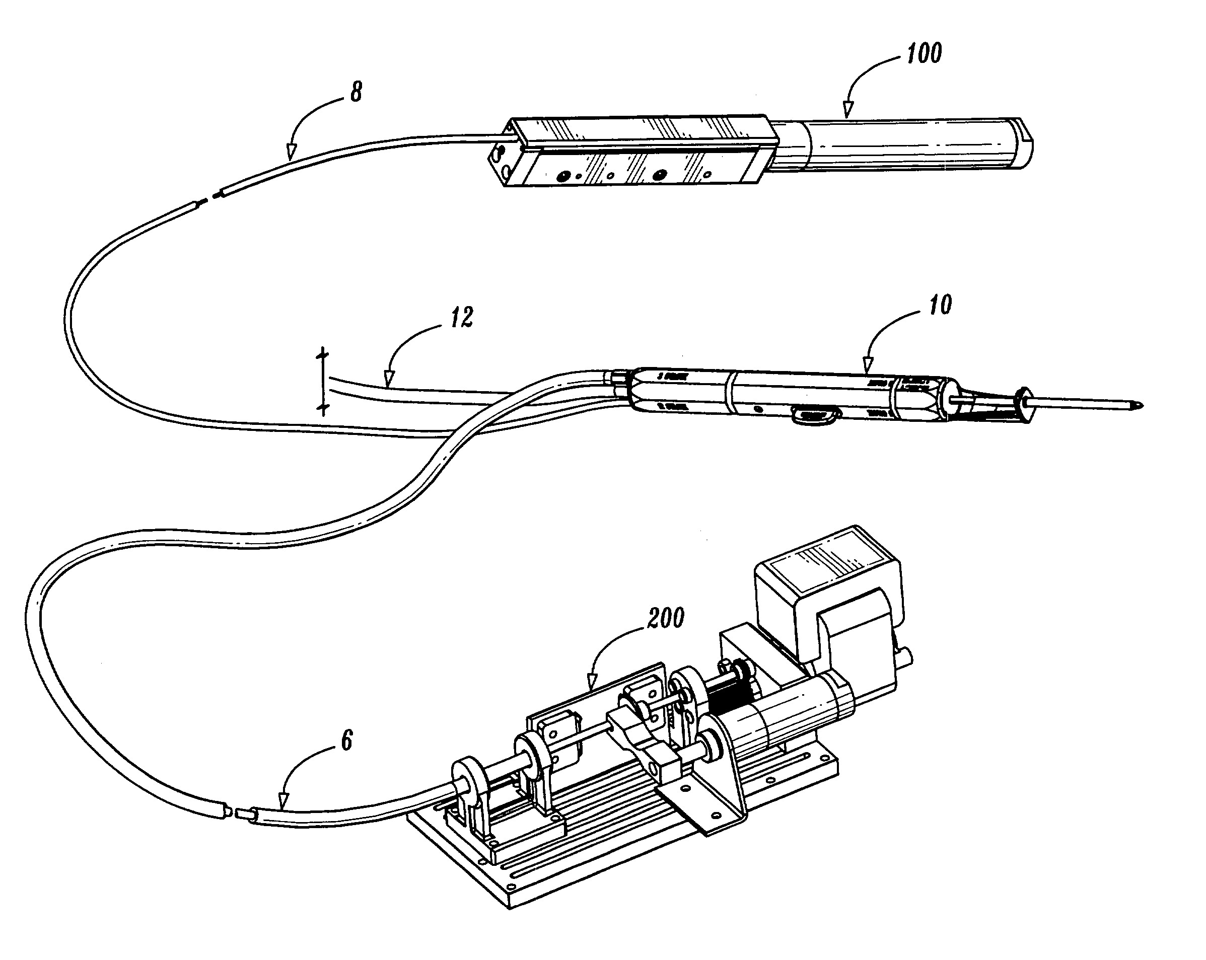
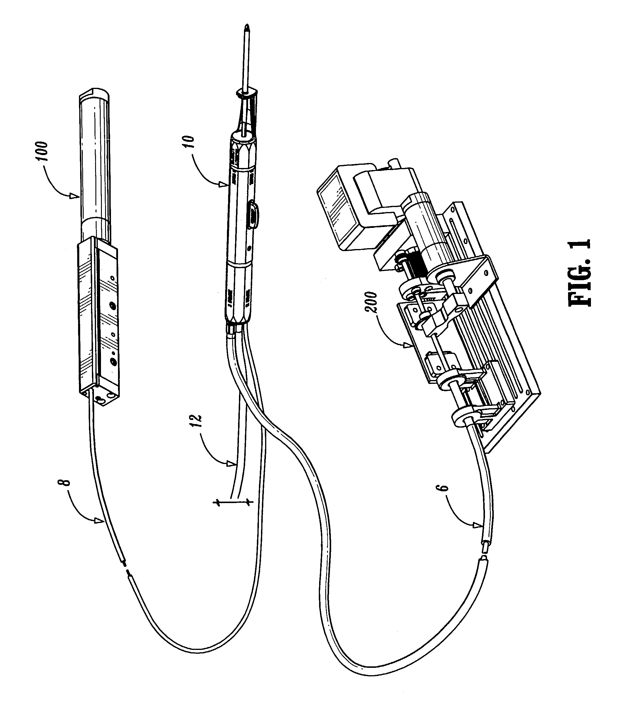
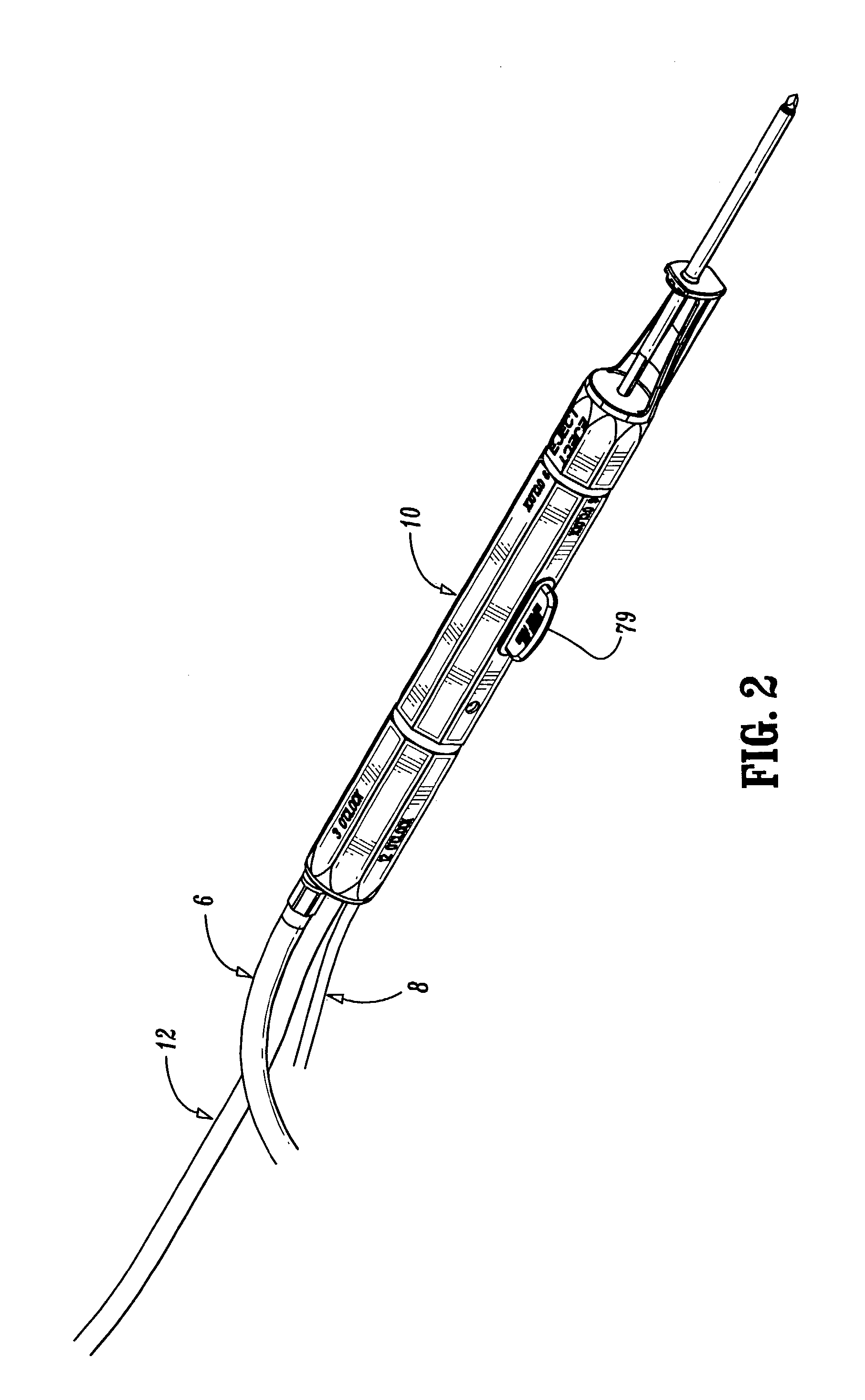
[0056]The presently disclosed biopsy system is illustrated in FIG. 1 and includes a single use loading unit (SULU) 10, a trocar driver 100 and a knife driver 200. A knife driver cable 6 extends between knife driver 200 and SULU 10, and a push / pull cable 8 extends between trocar driver 100 and SULU 10. Tr...


PUM


Abstract

A biopsy system for retrieving biopsy tissue samples from different regions of the body is disclosed. The biopsy system includes a single use loading unit having a trocar assembly and a knife assembly. A trocar driver is operably connected to the trocar assembly and is actuable to move a trocar between retracted and advanced positions. The trocar driver is disengaged from the trocar assembly prior to firing the trocar into a target tissue mass to reduce drag on the trocar during firing. A knife driver is operably connected to the knife assembly such that when actuated, a knife is both rotatably and axially advanced about the trocar.

Description

CROSS REFERENCE TO RELATED APPLICATIONS[0001]This application is a Continuation of U.S. patent application Ser. No. 09 / 659,468, filed on Sep. 11, 2000 now U.S. Pat. No. 6,712,773, the disclosure of which is hereby incorporated by reference herein.BACKGROUND[0002]1. Technical Field[0003]This disclosure relates to an apparatus and method for the biopsy of tissue specimens and, more particularly, to a single insertion, multiple sample percutaneous biopsy system and method of use.[0004]2. Background of Related Art[0005]It is often necessary to sample tissue in order to diagnose and treat patients suspected of having cancerous tumors, pre-malignant conditions and other diseases or disorders. Typically, in the case of suspected cancerous tissue, when the physician establishes by means of procedures such as palpation, x-ray or ultrasound imaging that suspicious conditions exist, a biopsy is performed to determine whether the cells are cancerous. Biopsy may be done by an open or percutaneou...

Claims


Application Information

Patent Timeline
Patent Type & Authority Patents(United States)
IPC IPC(8): A61B5/05A61B13/00A61B17/32A61B5/00A61B10/00A61B10/02
CPCA61B10/0275A61B2017/3409A61B2010/0208A61B10/0283
Inventor VIOLA, FRANK
Owner TYCO HEALTHCARE GRP LP
Why Eureka
Try Eureka