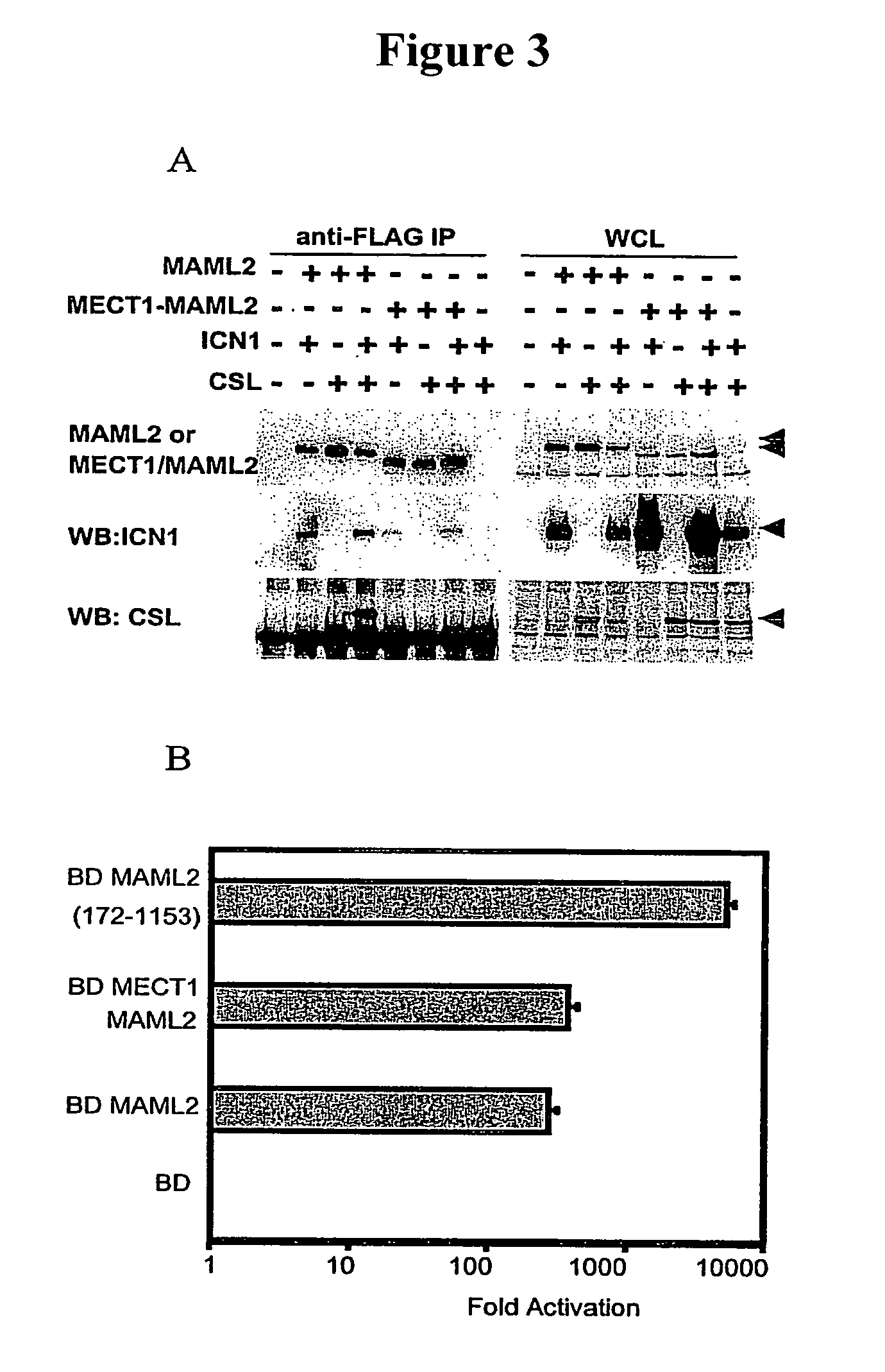
Detection of MECT1-MAML2 fusion products
a technology of fusion products and mectin, which is applied in the direction of instruments, biochemical equipment and processes, material analysis, etc., can solve the problems of incomplete excision, cancer represents a significant threat to human health, and salivary gland tumors can be deadly, so as to facilitate treatment regimens and improve pre- and/or post-operative management.
- Summary
- Abstract
- Description
- Claims
- Application Information
AI Technical Summary
Benefits of technology
Problems solved by technology
Method used
Image


Examples
example 1
Tumor Samples
[0129]In this Example, the tumor samples used during the development of the present invention are described. The H292 and H3118 (pulmonary MEC), the H727 (pulmonary carcinoid), and H2009 (non-small cell lung cancer) tumor cell lines were generated from patient biopsy samples at the National Naval Medical Center as known in the art (See, Carney et al., Cancer Res., 45:2913–23 [1985]; and Modi et al., Oncogene 19:4632–9 [2000]) using an IRB-approved tissue procurement protocol. The pulmonary MEC tumor sample (MECT #A) was obtained as an anonymous tumor sample approved for inclusion in this study by the NIH Office of Human Subjects Research. Human U2OS osteosarcoma cells were cultured in Dulbecco's modified Eagle's medium (DMEM) containing 10% Fetalclone I serum (HyClone). COS7 cells were cultured in RPMI 1640 medium supplemented with 10% FCS. NIH 3T3 cell transduced with the pBABE retrovirus encoding Jagged 2, or empty pBABE retrovirus, were maintained in DMEM containing ...
example 2
Spectral Karyotyping
[0130]In this Example, the spectral karyotyping methods used in the development of the present invention are briefly described (See, Tonon et al., Genes Chromosomes Cancer 27:418–23 [2000]). Specific chromosomes, kindly provided by Dr. Thomas Ried, were obtained by high-resolution flow sorting, and then amplified using two consecutive rounds of degenerate oligo-primed (DOP)-PCR amplification. Methods commonly used and widely known in the art were used for the spectral karyotyping experiments.
[0131]Spectrum Orange (Vysis), Rhodamine 110 (Perkin Elmer), and Texas Red (Molecular Probes) were used for the direct labeling of chromosomes, whereas Biotin-16-dUTP and Digoxigenin-11-DUTP (Roche) were used for the indirect labeling of chromosomes. After hybridization, biotin was detected with Avidin-Cy5 (Amersham) and digoxigenin-11-dUTP with mouse anti-digoxin (Sigma) followed by sheep anti-mouse antibodies custom-conjugated to Cy5.5 (Amersham). The slides were countersta...
example 3
Fluorescence in Situ Hybridization (FISH) Analysis
[0132]In this Example, the FISH analysis used during the development of the present invention is described. BAC clones were purchased from Research Genetics, Oakland BAC / PAC Resources, or provided by Dr. Raluca Jonescu (RP11-16K5). For the FISH analysis, BAC clones were labeled by nick translation. BAC clones RP11-676L3 and RP11-16K5 were used to identify translocations. Image acquisition was performed using a Sensys CCD camera (Photometrics), and Q-FISH software (Leica Microsystems Imaging Solutions). Using standard FISH protocols, specific translocation events were detected. Additional experiments to identify translocation using a single probe were also conducted using methods known in the art.
PUM
 Abstract
Description
Claims
Application Information