Ultrasonic method for visualizing brachytheraphy seeds

Active Publication Date: 2007-05-15
RIVERSIDE RES INST
View PDF11 Cites 10 Cited by
  • Summary
  • Abstract
  • Description
  • Claims
  • Application Information

AI Technical Summary

Benefits of technology

[0008]In accordance with a first embodiment of the invention there is provided a method for ultrasonically imaging implanted therapeutic seeds in tissue. The method includes determining acoustic reflection properties of the seeds in response to acoustic signals from an ultrasonic probe having selected signal characteristics. The selected signal characteristics may include a scanned beam to create two-dimensional images of the tissue and embedded seeds. The tissue and the seeds are insonated using acoustic signals having the selected signal characteristics and reflected acoustic signals are received from the tissue and the seeds. The reflected acoustic signals are analyzed using a correlation algorithm to generate an image of the tissue and the seeds with image portions corresponding to the seeds enhanced by the correlation algorithm.

Problems solved by technology

[Stock, et al, Int. J. Radiat. Oncol. Biol. Phys., 41:101–108, 1998] The conventional B-mode ultrasound images generated at the time of the procedure do not allow adequate visualization of the placed seeds because of clutter, shadowing and the loss of echo signals due to seed angulation.
Clutter often increases immediately during the procedure from hemorrhage and edema caused by the trauma of needle insertion.

Method used


Image

  • Ultrasonic method for visualizing brachytheraphy seeds
  • Ultrasonic method for visualizing brachytheraphy seeds
  • Ultrasonic method for visualizing brachytheraphy seeds

Examples


first embodiment

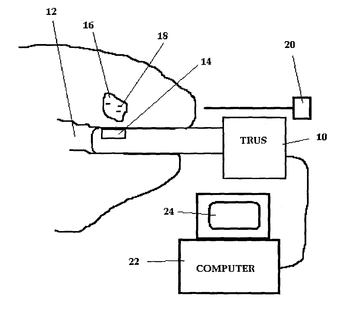
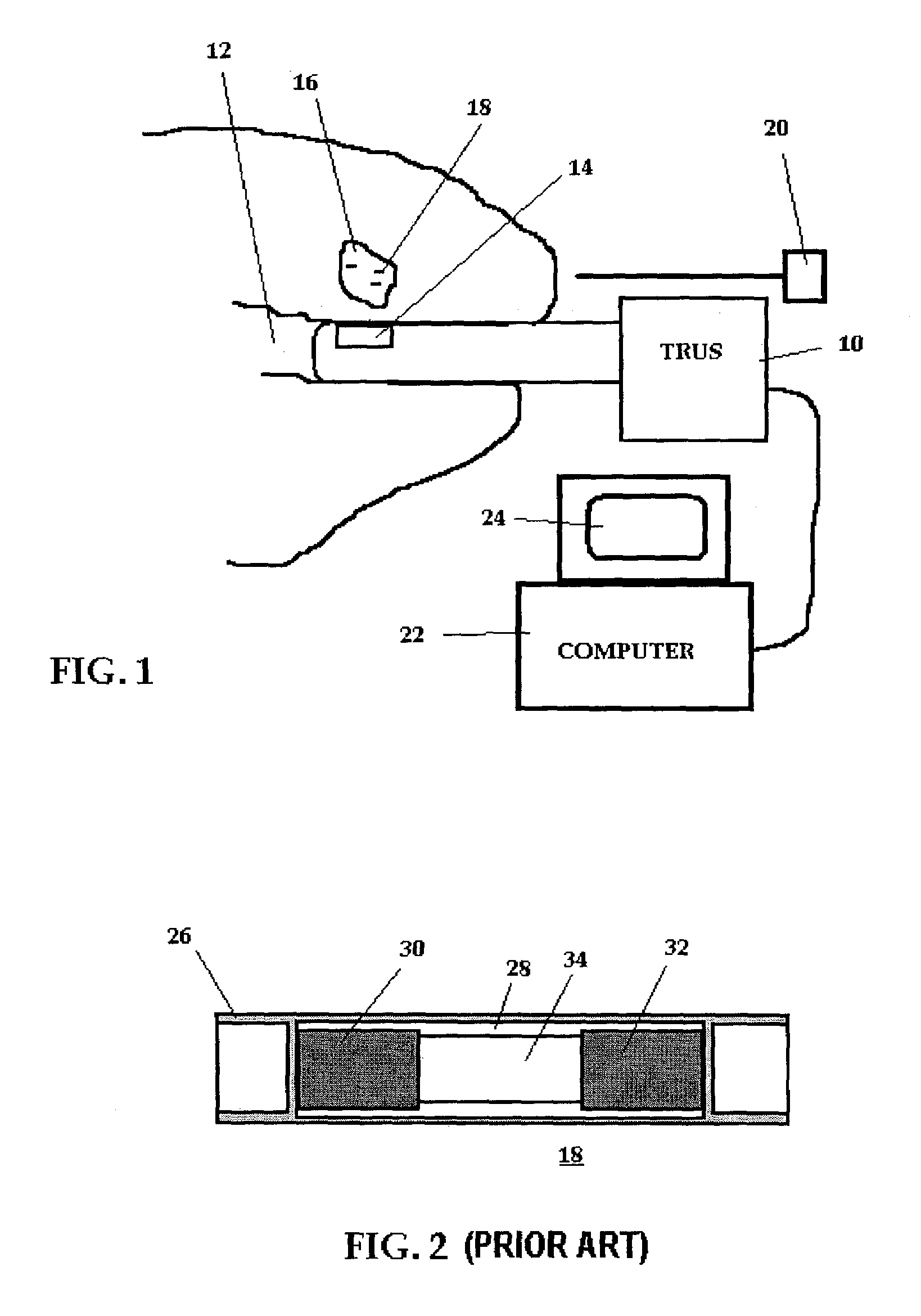
[0022]the method of the present invention will be described with reference to FIGS. 1 through 3. As used herein, the term therapeutic seeds includes brachytherapy seeds, which are well known, and other seeds of similar dimension which may be used for delivering a therapeutic agent to a localized region. Shown in FIG. 2 is a typical palladium seed 18 currently used in connection with brachytherapy of the prostate. The seed includes a thin cylindrical shell 26 of titanium, having overall dimensions of 4.5-mm length and 0.8-mm diameter. Different types of radioactive palladium and iodine seeds have different amounts of interior space and solid material. The illustrative palladium seed has graphite plugs 30, 32 at each end of the interior space 28, that is coated with radioactive palladium. The seed also contains a central lead plug 34, which is provided to enhance visibility on post-implantation CT scans. Iodine seeds come in a wide variety of configurations, but all seeds have the sam...

second embodiment

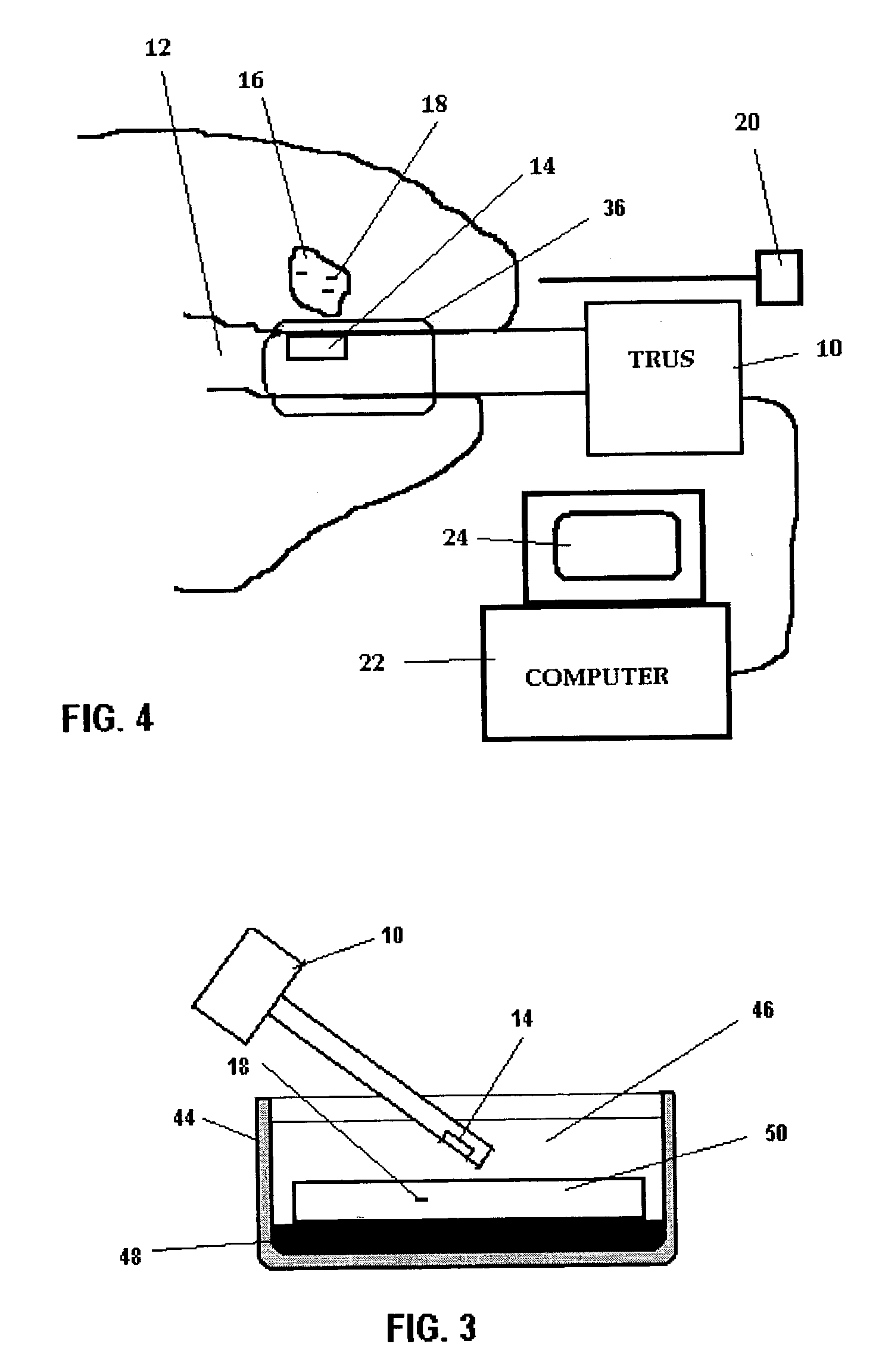
[0031]The second embodiment is based on cross-correlation analyses of echo signals from tissue before and after a high-strain deformation. In typical implementations of elastography applied to tissue, compression and deformation must be small in order to prevent decorrelation. In contrast, because the brachytherapy seeds are extremely stiff compared to tissue, they do not measurably distort upon compression, and large deformations beneficially decorrelate tissue signals, while retaining the correlation in signals from undeformed implanted seeds. At excessive strains, seeds may undergo complex motion, including out of plane motion and rotation, which can reduce correlation. The transducer axis is approximately perpendicular to the long axis of the seed. At least one RF frame is acquired before compression and one or more RF frames are acquired after each compression step. A one or two-dimensional correlation analysis between pre and post-deformation RF echo signals or their envelope ...


PUM


Abstract

Methods are provided for enhancing the image of brachytherapy seeds implanted in tissue using a transrectal ultrasound apparatus. In a first arrangement the resonance of the seeds is determined and the seeds are insonified using acoustic signals having a resonance frequency of the seeds to cause the seeds to vibrate. The vibrating seeds have enhanced detection using a Doppler ultrasound device. In a second arrangement the acoustic signature of the seeds is determined and used to enhance the imaging of the seeds using a correlation function. In a third arrangement images are taken of the tissue and implanted seeds prior to and during the application of mechanical stress to the tissue. A correlation between the unstressed and stressed tissue can cause enhanced imaging of the seeds.

Description

REFERENCE TO RELATED APPLICATIONS[0001]This application claims the benefit of U.S. Application Ser. No. 60 / 363,452, filed Mar. 12, 2002.BACKGROUND OF THE INVENTION[0002]The present invention was made in connection with a grant from the National Institute of Health Grant R21 CA88231[0003]Permanent interstitial implantation of iodine or palladium radioactive seeds is currently used for the treatment of prostate cancer. In connection with the implantation of such brachytherapy seeds a transrectal ultrasound (TRUS) examination of the prostate is used in the operating room. The TRUS probe is mounted on a fixture that contains a needle guidance template, which rests against the perineum and incorporates detents enabling the probe to be introduced into or withdrawn from the rectum in 5-mm steps. Scans are oriented in a transverse plane perpendicular to the probe axis. The fixture is advanced into the rectum until the seminal vesicles are imaged and scans are performed in planes separated b...

Claims


Application Information

Patent Timeline
IPC IPC(8): A61B8/00A61N5/00A61B8/08A61B8/12
CPCA61B5/0048A61B8/0833A61B8/0841A61B8/12A61B5/0051
Inventor ALAM, SHEIKH KAISARFELEPPA, ERNEST JOSEPHLIZZI, FREDERIC LOUIS
Owner RIVERSIDE RES INST
Why Eureka
Try Eureka