

Neutralizing human anti-IGFR antibody
a monoclonal, immune-like technology, applied in the direction of fungi, peptide/protein ingredients, unknown materials, etc., can solve the problems of low therapeutic value and limited human therapeutic utility
- Summary
- Abstract
- Description
- Claims
- Application Information
AI Technical Summary
Benefits of technology
Problems solved by technology
Method used
Image

Examples
example 1
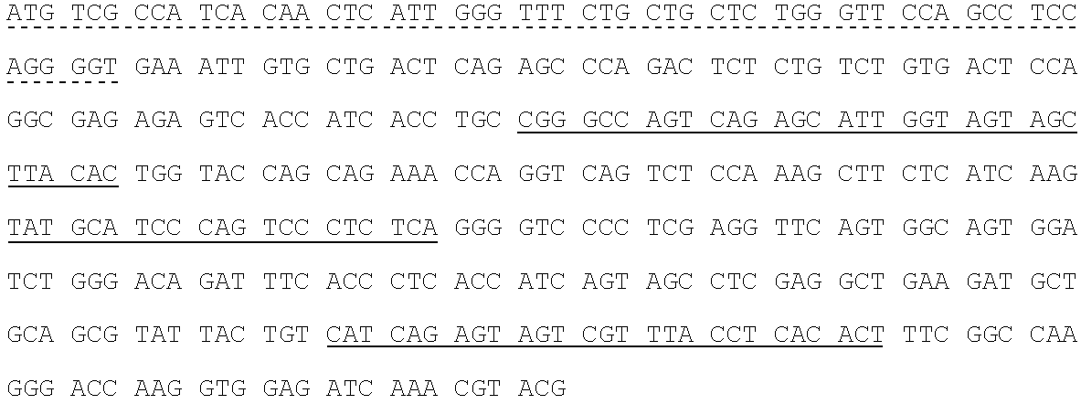
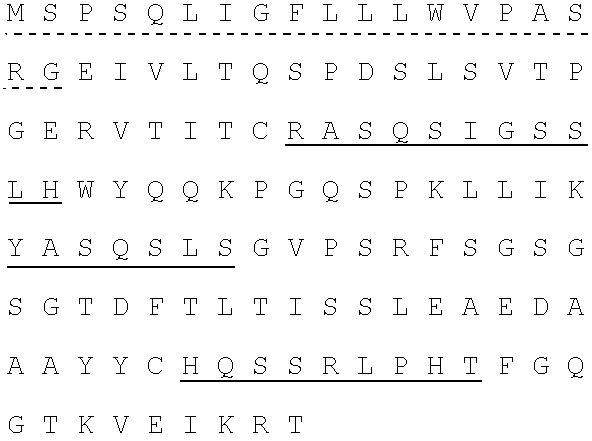
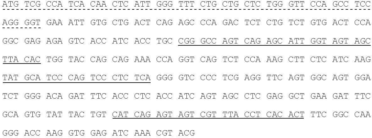
Construction of Fully Human Anti-IGFR1 Antibodies
1.0. Introduction.
[0250]Fully human monoclonal antibodies specific for human insulin-like growth factor receptor 1 (IGFR1) were generated from HuMab mice of the Hco7 genotype (see below), immunized with recombinant sIGFR1 and IGFR1 transfected HEK293 cells. A detailed description of Hco7 mice is provided in U.S. Pat. Nos. 5,545,806; 5,569,825; 5,625,126; 5,633,425; 5,661,016; 5,770,429; 5,789,650; 5,814,318; 5,874,299 and 5,877,397 and in Harding, et al., (1995) Ann. NY Acad. Sci. 764:536–546. Antibodies 1H3, 15H12 and 19D12 were isolated from a HuMab mouse (referred to herein as #23716) which was selected for fusion based on the presence of antigen specific serum IgG titers of 25,600 to the immunizing antigen. The 1H3, 15H12 and 19D121 antibodies were found to bind IGFR1.
2.0 Materials and Methods and Results.
[0251]2.1. Antigen.[0252]2.1.1. Mice were immunized with two forms of antigen: (1) live cells (IGFR1 transfected HEK293 cells) ...
example 2
Cell Based Receptor Binding Assay
[0411]A cell based receptor binding assay was used to determine if antibodies 1H3, 15H12 and 19D12 competed with IGF1 for binding to IGFR1.
[0412]In the assays, 96 well filter plates (1.2 μm pore) were pre-wet with 0.5% bovine serum albumin (BSA) / PBS for 2 hours at 4° C. The buffer was then removed with a vacuum manifold. Various concentrations of 6× control or test antibody (1H3, 15H12 or 19D12) were added to the wells (25 μL). The [125I]-IGF-1 ligand was then added to the wells at a final concentration of 0.375 nM in BSA / PBS. Cells were harvested with cell dissociation solution, counted with trypan blue, and resuspended in 0.5% BSA / PBS to a cell number of 1–3×10⁵ / ml. One hundred μl of cells (10,000–30,000) were added to each well. The plate was shaken at 4° C. for 1 hour. The plate was then aspirated and washed three times with ice cold PBS using a vacuum manifold. The filters were punched out and counted on a gamma counter. Data were analyzed for c...
example 3
IGFR1 Autophosphorylation Assay
[0414]The ability of 1H3, 15H12 and 19D12 to inhibit IGFR1 autophosphorylation was also determined.
[0415]Antibodies (1H3, 15H12 or 19D12) were added to cells bearing IGFR1 for various lengths of times. Cells were then stimulated with 10 ng / ml IGF-I for 5 min at 37° C. Cells were washed twice with cold PBS containing 0.1 mM sodium vanadate and lysed in lysis buffer (50 mM HEPES, pH 7.4, 150 mM NaCl, 10% glycerol, 1% Triton X-100, 1.5 mM MgCl2, protease inhibitors and 2 mM sodium vanadate). Lysates were incubated on ice for 30 min and then centrifuged at 13,000 RPM for 10 min at 4° C. Protein concentrations of the lysates were measured by a Coomassie colorimetric assay, and subjected to immunoprecipitation and Western blot analysis.
[0416]The results of these assays indicated that antibodies 1H3, 15H12 and 19D12 inhibited IGFR1 autophosphorylation with an IC50 of 0.10 nM.
PUM
| Property | Measurement | Unit |
|---|---|---|
| temperature | aaaaa | aaaaa |
| temperature | aaaaa | aaaaa |
| temperature | aaaaa | aaaaa |
Abstract
Description
Claims
Application Information