

Methods of determining polypeptide structure and function
a polypeptide and structure technology, applied in the field of methods of determining polypeptide structure and function, can solve the problems of difficult protein crystallization, complex structure determination by x-ray crystallography, large proteins (>40 kda) out of the range of structure determination by nmr techniques, etc., to inhibit or reduce activate or increase the activity of polypeptides
- Summary
- Abstract
- Description
- Claims
- Application Information
AI Technical Summary
Problems solved by technology
Method used
Image

Examples
example 1
Determination of a Ca2+-bound Active Site Structure of Calpain
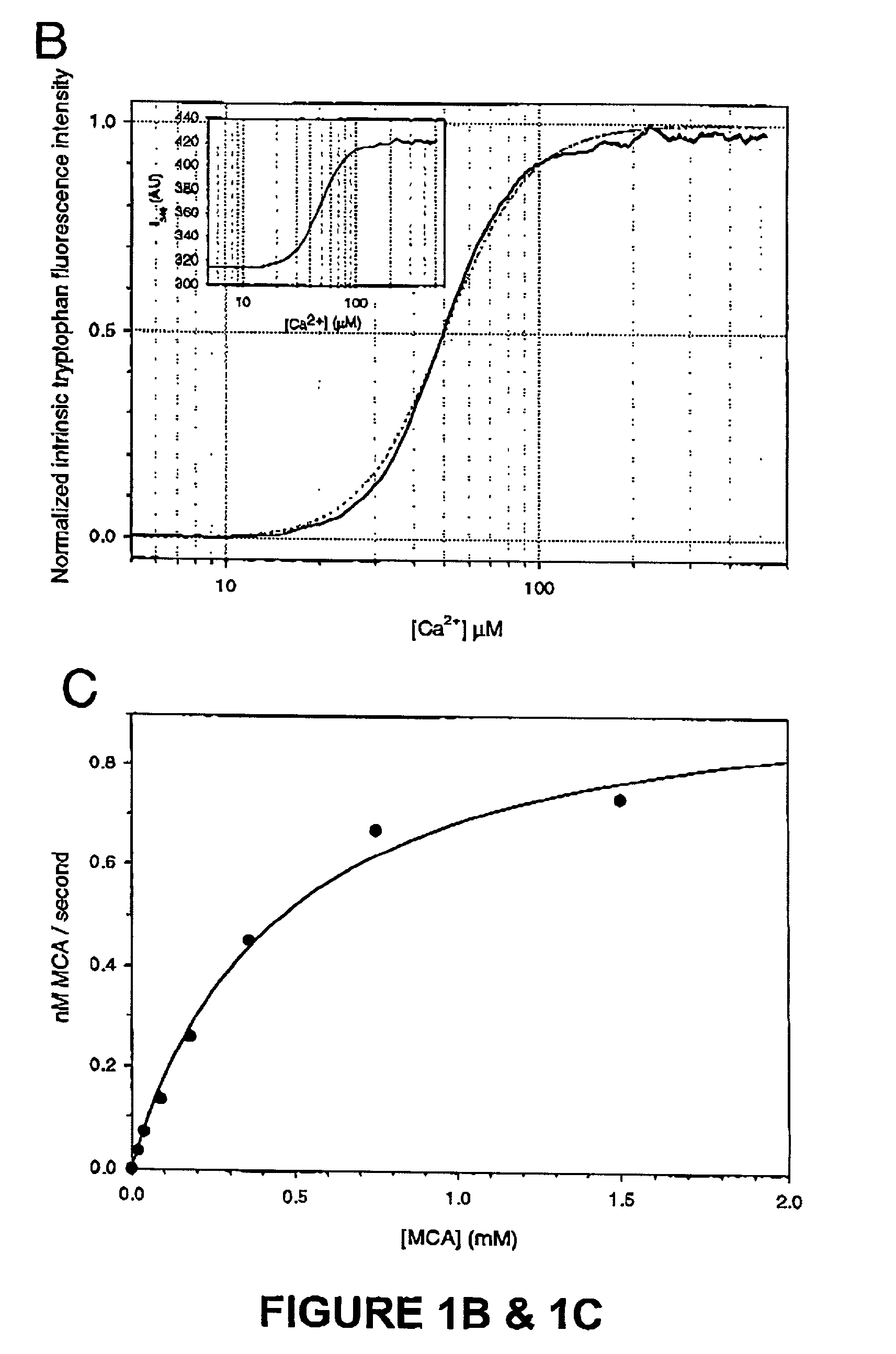
[0108]We have shown an evolutionarily conserved novel mechanism of calpain regulation by Ca2+. We have generated a structural entity containing the active site region of μ-calpain, which maintains the minimal requirements of a Ca2+ dependent cysteine protease. Ca2+-binds in a cooperative manner at two unique sites, one in each domain, resulting in a major conformational change that correlates with activation. The consequence of this conformational change is active site assembly with the catalytic residues arrangement closely resembling that of papain, as captured in the 2.1 Å crystal structure of μI-II. This structure supports the observed cooperativity between the Ca2+ sites suggesting a mechanism for activation of this protease. Our study provides the grounds for Ca2+ regulation of the large subunit homologues of the calpain super family. Moreover, the Ca2+-bound structure is a perfect template for active site-directed ...
example 2
μI-II Binds Ca2+ and is a Ca2+-dependent Cysteine Protease
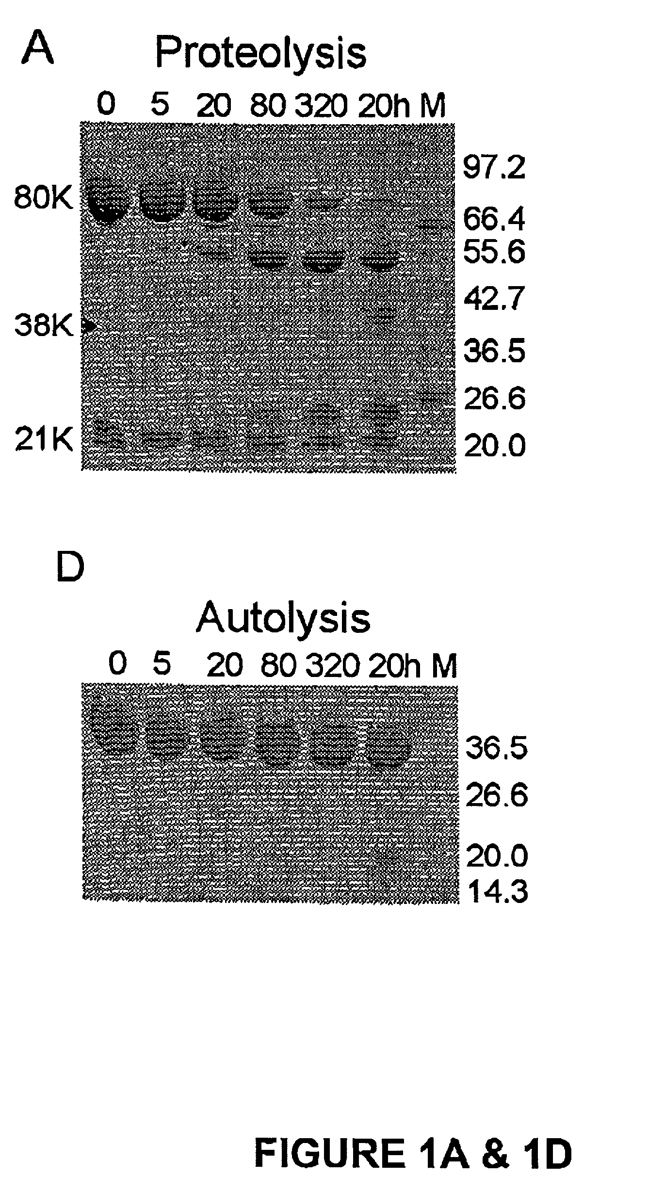
[0116]The active site of m-calpain in the absence of Ca2+ is not formed as shown in the rat (Hosfield, C. M., et al, EMBO J. 18:6880–6889 (1999)) and human (Strobl, S., et al., Proc. Natl. Acad. Sci. U.S.A. 97:588–592 (2000)) structures. From examining the inactive structures it is evident that well-defined interactions on either side of the active site domains I and II could keep the active site domains apart. The independent expression of the active site domains I+II was therefore expected to be free of destabilizing interactions from neighboring regions and perhaps show activity. We undertook this experiment and to our surprise the construct containing solely the active site of μ-calpain, μI-II was inactive; that is in the absence of Ca2+. The μI-II construct extends from the second calpain autolysis site, residue 29, which defines the start of domain I, to the end of domain II (residue 356). Its domain boundaries were cho...
example 3
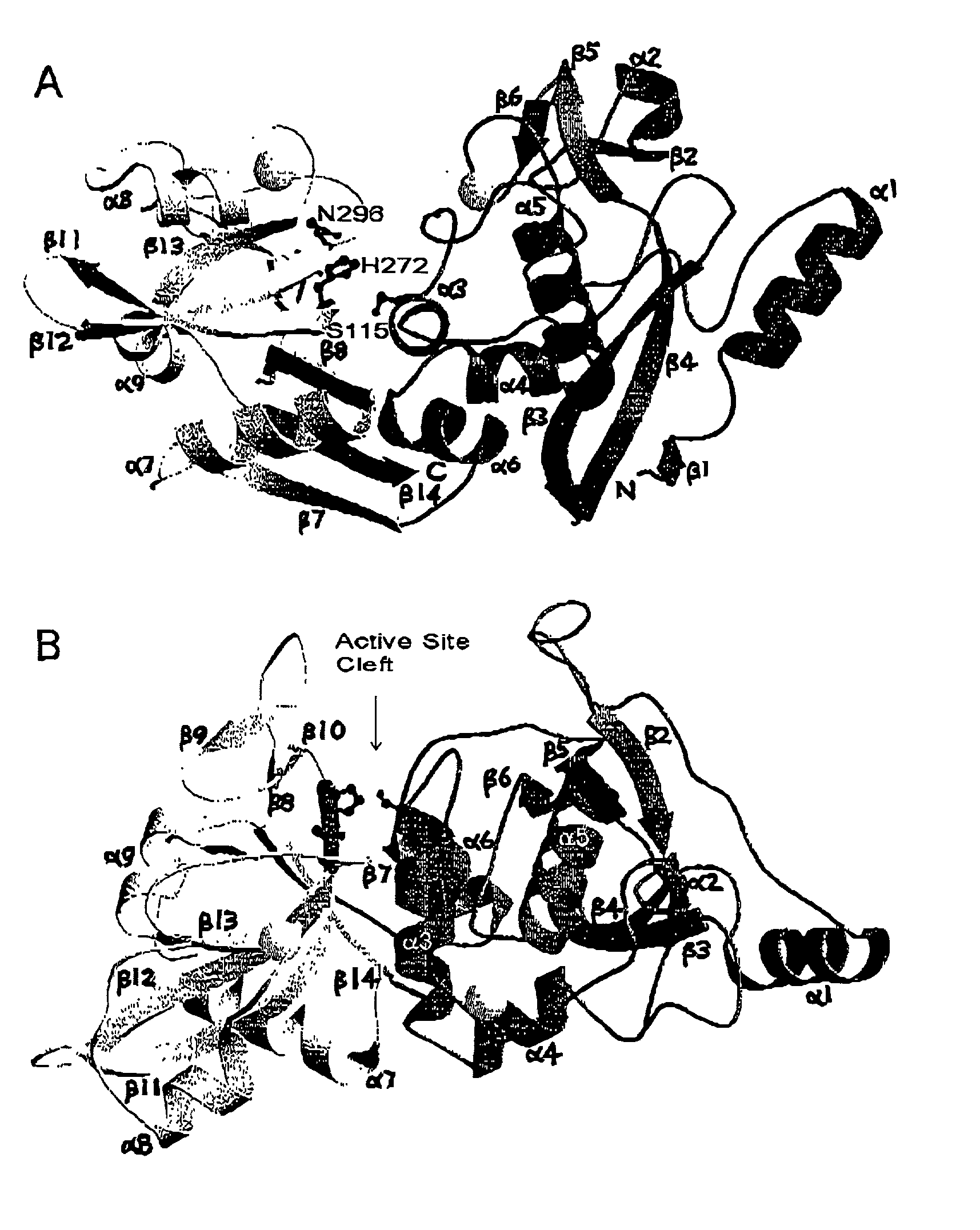
Overall Structure
[0121]Over most of the structure, the Ca2+-bound structure of μI-II is very similar to that of domains I and II from the inactive rat (Hosfield, C. M., et al., EMBO J 18:6880–6889 (1999)) or human (Strobl, S., et al., Proc. Natl. Acad. Sci. U.S.A. 97:588–592 (2000)) m-calpain heterodimer. The secondary structure elements and their arrangement as observed in the inactive human structure match very closely those of the μI-II structure (FIG. 2). Domain I maintains the core α-helix (α5) surrounded by two β-sheets on one side and a cluster of α-helices on the other side. In addition to the two antiparallel β-sheets that form the core of domain II in the inactive structure, a new antiparallel β-sheet is formed between the short strands β9 and β10 (FIG. 2B) which are not interacting in the inactive structure. The two most significant structural differences between the active and inactive forms are the presence of two Ca2+ ions, one bound at each domain, and the difference ...
PUM
| Property | Measurement | Unit |
|---|---|---|
| wavelength | aaaaa | aaaaa |
| a wavelength | aaaaa | aaaaa |
| wavelength | aaaaa | aaaaa |
Abstract
Description
Claims
Application Information