Methods for modulating angiogenesis via VEGF
a technology of angiogenesis and vegf, which is applied in the direction of angiogenin, antibody medical ingredients, peptide/protein ingredients, etc., can solve the problems of further harming patients, and achieve the effect of stimulating or inhibiting angiogenesis
- Summary
- Abstract
- Description
- Claims
- Application Information
AI Technical Summary
Benefits of technology
Problems solved by technology
Method used
Image
Examples
example 1
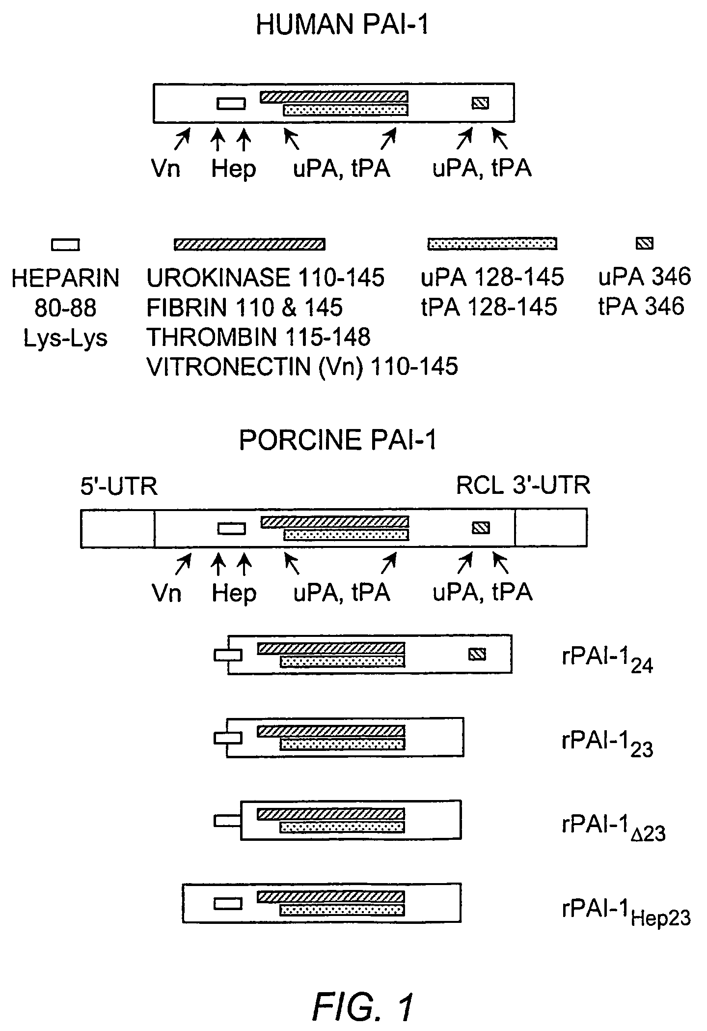
PAI-1 Gene Deletions
[0092]The DNA encoding the truncated PAI-1 proteins was obtained by deleting the porcine PAI-1 gene (poPAI-1) (Bijnens, et al. (1997) Thromb. Haemost. 77:350–356; Accession number Y11347); SEQ ID NO:9). The selection of gene fragments was based on the poPAI-1 sequences that correspond to the human PAI-1 gene (huPAI-1) (Bosma, et al. (1988) J. Biol. Chem. 263:9129–9141; Accession number J03764; SEQ ID NO:10) sequence reported to code for functional domains in human PAI-1 (Reilly and Hutzelmann (1992) supra). Each DNA fragment was isolated from porcine aortic endothelial cells by reverse transcribing RNA into cDNA. The cDNA was made double-stranded in a polymerase chain reaction (PCR) containing porcine PAI-1-specific primers. Primers for rPAI-123 amplification are known, for example, Mulligan-Kehoe, et al. ((2001) supra). Primers used to PCR amplify the cDNA into double-stranded DNA coding for rPAI-1Δ23 are 5′ primer: 5′-GGAATTCATGGATGAGATCAGCACGG-3′ (SEQ ID NO:3)...
example 2
Anti-Angiogenic Effects of rPAI-1 Proteins
[0093]Characterization of the rPAI-1 Protein Interactions with heparin, uPA, and Plasminogen. The functionality of the rPAI-1 proteins was first tested by incubating each truncated protein with uPA and plasminogen to assess the proteolytic activity of the products of the reaction, as known in the art, for example Mulligan-Kehoe, et al. ((2001) supra). The functionality of each rPAI-1 isoform was then tested in a reaction with heparin bound to Sepharose beads. First, the rPAI-123, rPAI-1Δ23, rPAI-1Hep23, and rPAI-124 proteins (20 μg) were each incubated with 50 μl of heparin-bound Sepharose beads (Amersham Pharmacia Biotech, Piscataway, N.J.) for 2 hours at 37° C. The unbound protein was separated from the heparin-Sepharose-bound protein complex by microcentrifugation at 4° C. for 15 minutes. The proteins bound to the Sepharose beads were washed in TBS / Tween-20 followed by TBS washes. Bound samples were reacted with uPA (0.5 IU / 1 μg or rPAI-1...
example 3
VEGF-A Interactions
[0104]Biochemical Interactions of a VEGF-Heparin Complex with rPAI Proteins, uPA, and Plasmin. VEGF was isolated from bovine aortic endothelial cells by first incubating the cells overnight in serum-free DMEM. The medium was changed before adding 100 μg / ml heparin (Sigma, St. Louis, Mo.) for 4 hours at 37° C. The serum-free DMEM containing the VEGF-heparin complex was precipitated in 80% ethanol. The serum-free medium from which the VEGF-heparin complex was isolated, was probed for VEGF-A121 (Santa Cruz Biotechnology, Inc., Santa Cruz, Calif.) to insure that his non-heparin-binding VEGF isoform did not precipitate with the VEGF-heparin complex. VEGF-A121 was not detected. The heparin-VEGF complex was incubated with rPAI-123, rPAI-1Δ23, rPAI-1Hep23, or rPAI-124 protein (3, 15, and 30 nM) for 2 hours at 37° C. Either uPA (0.25 IU) or plasmin (made from 1.0 IU plasminogen and 0.25 IU uPA) was added to the VEGF-heparin-rPAI-1 reaction. After an additional 2 hours incu...
PUM
| Property | Measurement | Unit |
|---|---|---|
| molecular mass | aaaaa | aaaaa |
| molecular mass | aaaaa | aaaaa |
| molecular mass | aaaaa | aaaaa |
Abstract
Description
Claims
Application Information
 - R&D
- Intellectual Property
- Life Sciences
- Materials
- Tech Scout
- Unparalleled Data Quality
- Higher Quality Content
- 60% Fewer Hallucinations
Browse by: Latest US Patents, China's latest patents, Technical Efficacy Thesaurus, Application Domain, Technology Topic, Popular Technical Reports.
© 2025 PatSnap. All rights reserved.Legal|Privacy policy|Modern Slavery Act Transparency Statement|Sitemap|About US| Contact US: help@patsnap.com

