
Magnetic resonance cylindrical body coil and method for generating a homogeneous RF field

a cylindrical body coil and magnetic resonance technology, applied in the field of magnetic resonance apparatus, can solve the problems of image artifacts, large amount of energy radiated into the patient, time-consuming excitation sequence, etc., and achieve the effect of minimizing field formation, maximum homogeneity, and simple and fast generation

Active Publication Date: 2007-07-10
SIEMENS HEALTHCARE GMBH
View PDF6 Cites 12 Cited by
  • Summary
  • Abstract
  • Description
  • Claims
  • Application Information

AI Technical Summary

Benefits of technology

[0014]Because the RF field is also generated or excited only where the image should be acquired, or where image information should be read out, in the inventive magnetic resonance apparatus as a consequence of the defined, locally-delimited excitation field generation, the use of the saturation pulse required in the prior art is no longer necessary. The sequence workflow consequently can be accelerated and the specific absorption rate (SAR) is inevitably reduced because the saturation pulse is not emitted. The occurrence of artifacts that can also result as described from a rephasing in the prior art is advantageously avoided.
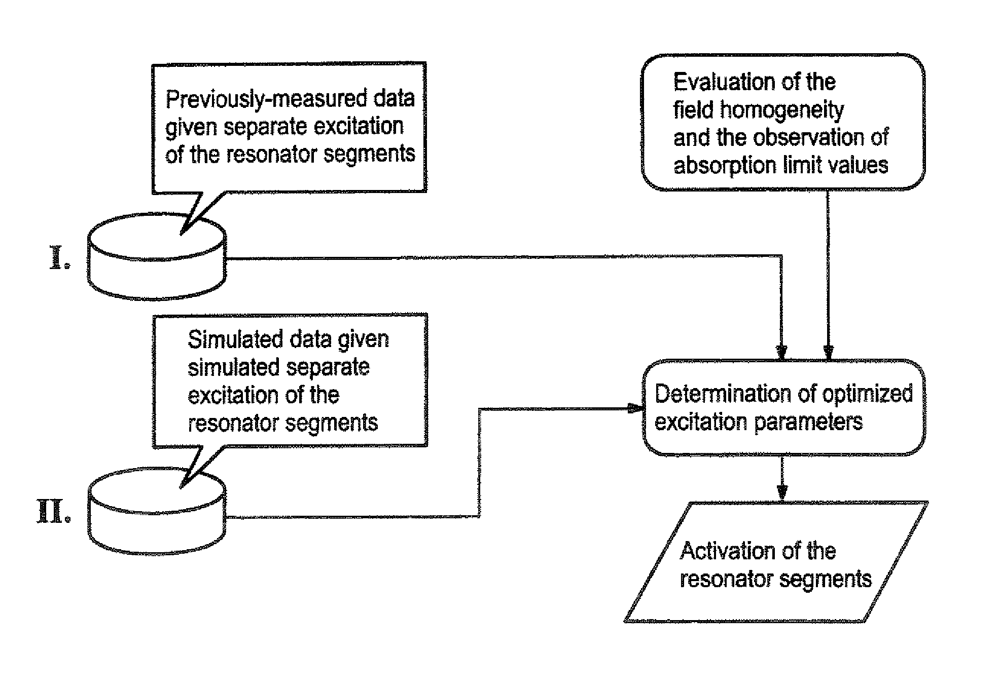
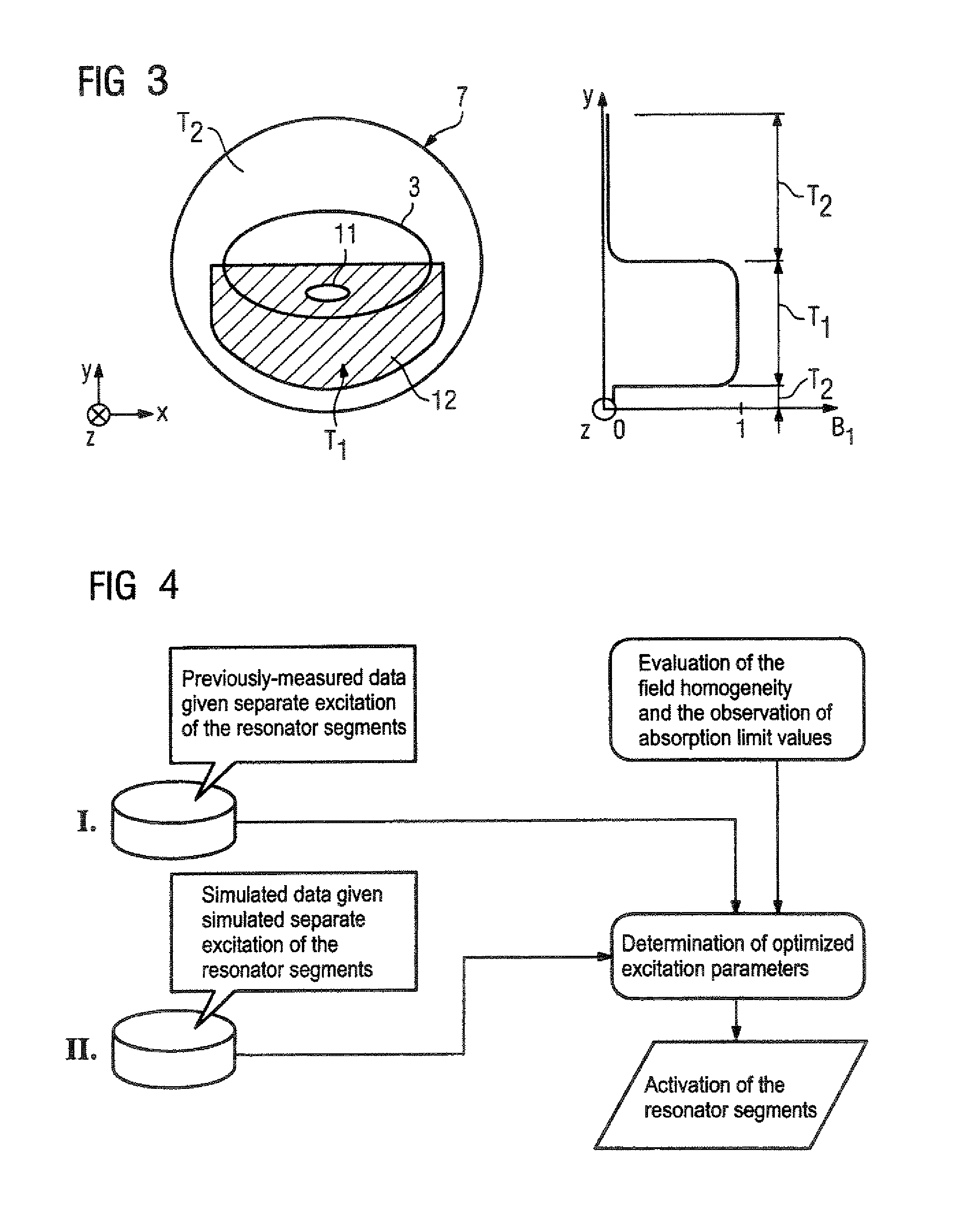
[0019]On the basis of such a procedure it is possible (if applicable, with repetition of the steps of the computational superimposition, the homogeneity evaluation and the determination of the parameters (iterative optimization)) to fashion or to shape the RF field corresponding to the clinical question, thus corresponding to the actual examination volume to be acquired, and to simultaneously minimize the field formation in the second sub-volume. For example, if the excitation field for a spinal column acquisition should be generated only in the lower region of the coil volume, such that the abdominal wall is not situated in the excitation field, the described parameter determination ensues such that just this field geometry is set, by excitation of a few or all resonator segments. For each resonator segment, the corresponding excitation parameters (starting from a normal excitation as a basis of, for example, the iterative parameter prioritization) are determined that cause the individual fields to be generated such that the overall field distribution is adjusted, such that only the desired sub-volume is excited, or the excitation field is generated only in the desired sub-volume, while the other volume region is essentially field-free. Using a changed, optimized set of excitation parameters, a subject-specific excitation of the resonator segments that is thus dependent on the examination subject can ensue for generation of a locally-optimized, geometrically optimally-shaped, polarized magnetic field with maximum homogeneity. Using such a procedure, the inventive magnetic resonance apparatus and method allow a simple and fast generation of an excitation field with maximum homogeneity in the examination volume, defined by the excitation field sub-volume. Complete scanning by simultaneous or time-offset grouped or individualized activation of only the resonator segments necessary to acquire the individual fields is required for this purpose. A diagnostic patient exposure is not needed. The parameters required for the subsequent diagnostic image acquisition can be determined in a simple and fast manner. Since the evaluation algorithm serves for the optimization of the excitation field with regard to its homogeneity, the image acquisition can ensue on the basis of an optimally homogeneous excitation field, such that the maximum information yield is possible from the limited examination volume without inhomogeneities of the image quality resulting from the non-excitation of other volume regions that are not of interest.

Problems solved by technology

A further circumstance that can lead to image artifacts is fold-over artifacts in the phase encoding direction when, for time reasons, the entire excited imaging region is not to be acquired with a sufficiently dense sampling of k-space.
This excitation sequence is time-consuming because two separate pulses must be provided.
Moreover, a relatively large amount of energy is radiated into the patient, which increases the specific absorption rate SAR, which is associated with limitations with regard to the number of slices, the repetition time or the measurement time.
The problem also exists that the dephased magnetization may possibly be re-phased with the subsequent excitation pulse, such that in spite of the saturation pulse it can contribute to the image information as an image artifact.

Method used


Image

  • Magnetic resonance cylindrical body coil and method for generating a homogeneous RF field
  • Magnetic resonance cylindrical body coil and method for generating a homogeneous RF field
  • Magnetic resonance cylindrical body coil and method for generating a homogeneous RF field

Examples


Embodiment Construction

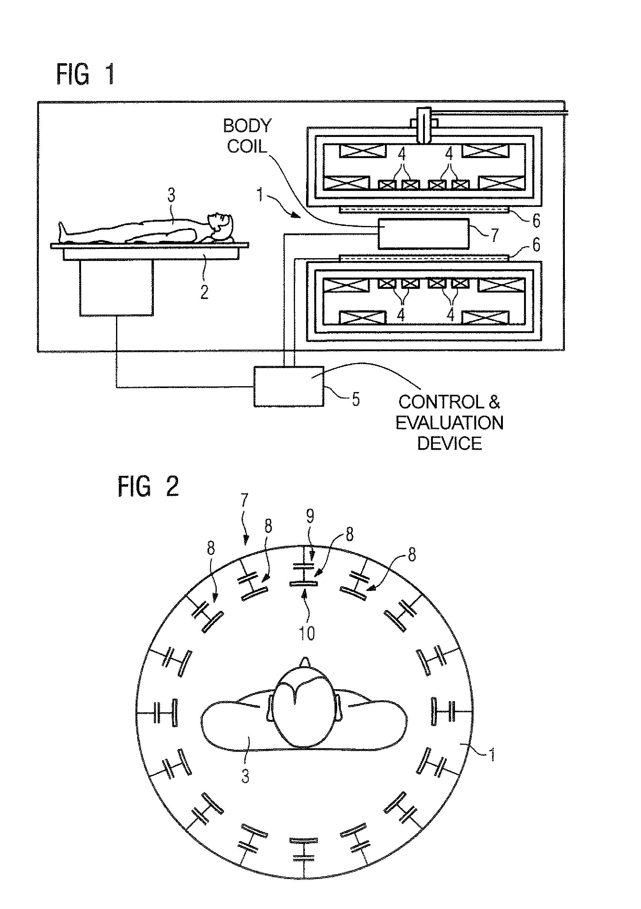
[0029]FIG. 1 shows an inventive magnetic resonance examination system that has an examination region 1. An examination subject 3 (here a person) can be introduced into the examination region 1 by means of a patient bed 2. The examination region 1 that corresponds to the examination volume is charged with a basic magnetic field by means of a basic field magnet 4. The basic magnetic field is temporally constant (static) and spatially as homogeneous as possible. It exhibits a magnetic field strength that is advantageously 3 T or more.

[0030]The basic field magnet 4 can be fashioned as a superconductor. No further activations by a control and evaluation device 5 (via which the system operation is controlled) are thus necessary.

[0031]The magnetic resonance system also has a gradient system 6 by means of which the examination region 1 can be charged with gradient magnetic fields. The gradient system 6 can be activated by the control and evaluation device 5, such that gradient currents flow...


PUM


Abstract

A magnetic resonance apparatus has a cylindrical body coil composed of a number of resonator segments distributed around the circumference and a control device for separate activation of the individual resonator segments. The resonator segments are electromagnetically decoupled from one another. The activation generates a radio-frequency magnetic field inside the coil for spin excitation in an examination volume. The resonator segments are activated such that the excitation field is generated only in at least one first sub-volume forming the examination volume, and at least one second sub-volume, that is not to be excited, is essentially free of the excitation field.

Description

BACKGROUND OF THE INVENTION[0001]1. Field of the Invention[0002]The present invention concerns a magnetic resonance apparatus of the type having a cylindrical body coil formed by a number of resonator segments distributed around the circumference and a control device for separately activating of the individual resonator segments (the resonator segments being electromagnetically decoupled from one another) so as to generate a radio-frequency field inside the coil for magnetic resonance (MR) spin excitation in an examination volume.[0003]2. Description of the Prior Art[0004]Magnetic resonance tomography is one of the imaging methods in medical diagnostics that uses the interaction of an external field (here a magnetic field) with the human body for imaging. Known magnetic resonance apparatuses have a (normally) cylindrical body coil that is formed by a number of resonator segments distributed around the circumference. Each resonator segment normally is formed of at least one capacitor...

Claims


Application Information

Patent Timeline
Patent Type & Authority Patents(United States)
IPC IPC(8): G01V3/00
CPCG01R33/34046G01R33/3607
Inventor FEIWEIER, THORSTEN
Owner SIEMENS HEALTHCARE GMBH
Learn More