
System and method for detecting cardiac ischemia using an implantable medical device

a medical device and implantable technology, applied in the field of implantable medical devices, can solve the problems of tissue death, cardiac tissue beyond the blockage no longer receiving oxygen, and a substantial portion of heart muscle ceases to function, and achieves the effect of saving power consumption, cost and other factors

Inactive Publication Date: 2007-09-25
PACESETTER INC
View PDF54 Cites 28 Cited by
  • Summary
  • Abstract
  • Description
  • Claims
  • Application Information

AI Technical Summary

Benefits of technology

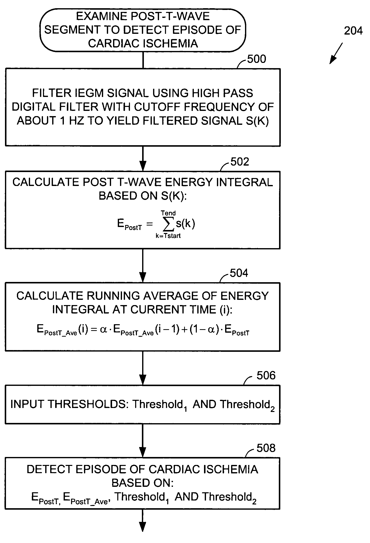
[0010]In accordance with the invention, a technique is provided for use with an implantable medical device for detecting cardiac ischemia. Electrical cardiac signals are received and segments of the cardiac signals subsequent to ventricular repolarization are identified (i.e. post-T-wave segments). Cardiac ischemia is then detected based on an examination of the post-T-wave segments. By detecting cardiac ischemia based on post-T-wave signals, the problems noted above arising when trying to exploit ST segments are substantially overcome. In particular, as will be explained, a highpass filter having a relatively high cutoff frequency may be used, thus eliminating the need to use a low cutoff frequency filter, which typically requires a large capacitor. In an exemplary embodiment, upon detecting of the onset of an episode of cardiac ischemia, warning signals are generated, which include both a “tickle warning” applied to subcutaneous tissue and a short range telemetry warning signal transmitted to a device external to the patient. Once the tickle warning is felt, the patient positions an external warning device above the chest, which receives the short range telemetry signals and provides audible or visual verification of the ischemia warning. In this manner, the warning is reliably relayed to the patient, without requiring a long range telemetry system and so savings are achieved in power consumption, cost and other factors.

Problems solved by technology

With AMI, a substantial portion of heart muscle ceases to function because it no longer receives oxygen, usually due to significant blockage of the coronary artery.
Eventually, the blood clot completely blocks the coronary artery and so heart tissue beyond the blockage no longer receives oxygen and the tissue dies.
In many cases, an AMI proves fatal because too much tissue is damaged to allow continued functioning of the heart muscle.
Indeed, AMI is a leading cause of death in the United States and worldwide.
In other cases, although the AMI itself may not be fatal, it strikes while the victim is engaged in potentially dangerous activities, such as driving vehicles or flying airplanes, and the severe pain and possible loss of consciousness associated with AMI results in serious accidents.
Even if the victim survives the AMI, quality of life may be severely restricted thereafter, because the heart is unable to adequately function due to regions of dead tissue.
Episodes of cardiac ischemia may also trigger certain types of arrhythmias that may prove fatal, particularly ventricular fibrillation (VF) wherein the ventricles of the heart beat chaotically resulting in little or no net flow of blood from the heart to the brain and other organs.
Problems, however, arise when attempting to detect cardiac ischemia using ST segments.
Unfortunately, highpass filters requiring low cutoff frequencies are not well-suited for use within implantable medical devices.
In particular, such filters require very large capacitors, which add significantly to the size and weight of the implantable device.
In addition, because the ST segment primarily consists of low-frequency signals, techniques based upon an analysis of those segments may not be particularly reliable.
Long range telemetry devices require a considerable amount of power and can add significantly to the size, weight and cost of the implantable device.
Short range telemetry devices may not be sufficient to ensure that the warning is received.

Method used


Image

  • System and method for detecting cardiac ischemia using an implantable medical device
  • System and method for detecting cardiac ischemia using an implantable medical device
  • System and method for detecting cardiac ischemia using an implantable medical device

Examples


Embodiment Construction

[0028]The following description includes the best mode presently contemplated for practicing the invention. The description is not to be taken in a limiting sense but is made merely for the purpose of describing the general principles of the invention. The scope of the invention should be ascertained with reference to the issued claims. In the description of the invention that follows, like numerals or reference designators will be used to refer to like parts or elements throughout.

[0029]Overview of Implantable Device

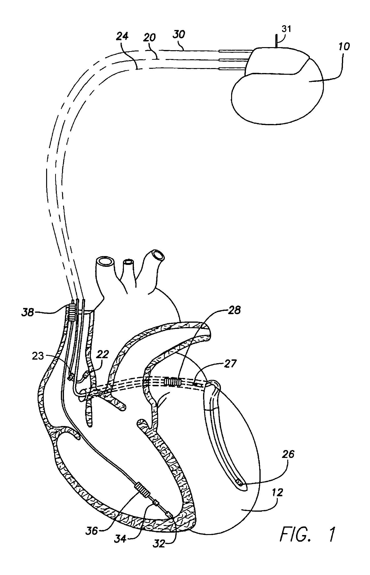
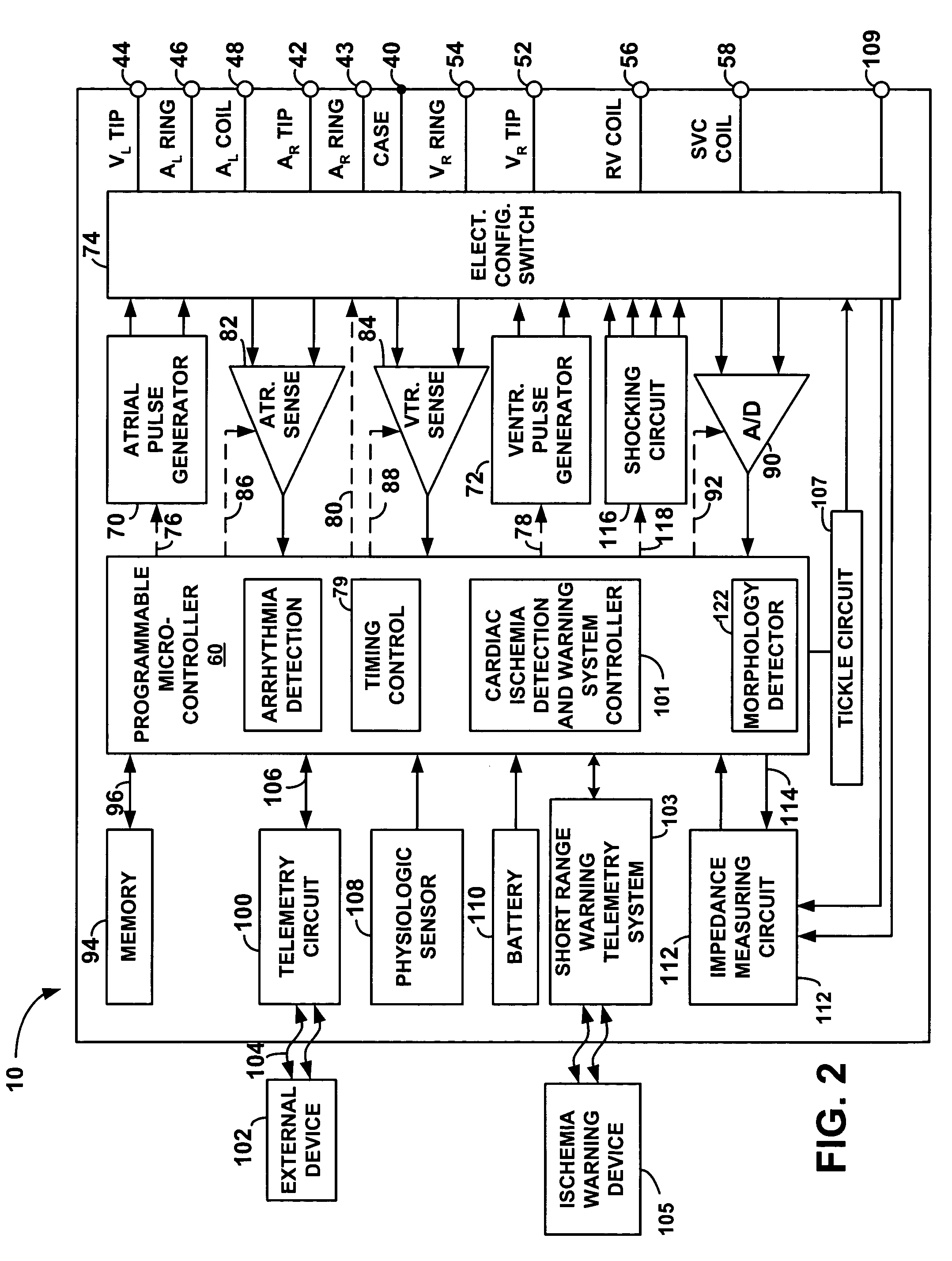
[0030]As shown in FIG. 1, there is a stimulation device 10 in electrical communication with the heart 12 of a patient by way of three leads, 20, 24 and 30, suitable for delivering multi-chamber stimulation and shock therapy. To sense atrial cardiac signals and to provide right atrial chamber stimulation therapy, the stimulation device 10 is coupled to an implantable right atrial lead 20 having at least an atrial tip electrode 22, which typically is implanted in the righ...


PUM


Abstract

A technique is provided for detecting episodes of cardiac ischemia based on an examination of post-T-wave signal segments. Since cardiac ischemia is often a precursor to acute myocardial infarction (AMI) or ventricular fibrillation (VF), the technique thereby provides a method for predicting the possible onset of AMI or VF so that a warning may be delivered to the patient. The warning preferably includes both a perceptible electrical notification signal applied directly to subcutaneous tissue and a warning signal delivered via short range telemetry to a handheld warning device external to the patient. In one example, the onset of cardiac ischemia is identified by detecting a sharp falling edge within post-T-wave signals by filtering the signals using a high-pass filter having a cutoff frequency of at least 1 Hz. The total amount of energy in the filtered signal is calculated and compared against various thresholds.

Description

CROSS REFERENCE TO RELATED APPLICATIONS[0001]This application is related to copending U.S. patent applications:[0002]1) Ser. No. 10 / 603,398, filed Jun. 24, 2003, titled “System and Method for Detecting Cardiac Ischemia Based on T-Waves Using an Implantable Medical Device”; and 2) Ser. No. 10 / 606,299, filed Jun. 24, 2003, titled “System and Method for Detecting Cardiac Ischemia Based on T-Waves Using an Implantable Medical Device,” which are incorporated herein by reference.FIELD OF THE INVENTION[0003]The invention generally relates to implantable medical devices such as pacemakers and implantable cardioverter / defibrillators (ICDs) and, in particular, to techniques for detecting cardiac ischemia.BACKGROUND OF THE INVENTION[0004]Cardiac ischemia is a condition whereby heart tissue does not receive adequate amounts of oxygen and is usually caused by a blockage of an artery leading to the heart tissue. If sufficiently severe, the ischemia results in an acute myocardial infarction (AMI),...

Claims


Application Information

Patent Timeline
IPC IPC(8): A61B5/0452
CPCA61B5/0452A61B5/352A61B5/349
Inventor WANG, ZIFEIFARD, MOHSSENSNELL, JEFFERY D.
Owner PACESETTER INC
Learn More