Method for radiation treatment

a radiation treatment and radiation therapy technology, applied in the field of breast cancer treatment, can solve the problems of inability to move the patient for imaging, logistically difficult if not impossible, and the intraoperative radiation treatment is difficult if not impossible, and achieve the effect of accurately treating the prescribed volume without damage to sensitive areas

Inactive Publication Date: 2008-01-29
XOFT INC
View PDF4 Cites 70 Cited by
  • Summary
  • Abstract
  • Description
  • Claims
  • Application Information

AI Technical Summary

Benefits of technology

[0016]In one preferred procedure, the ionizing radiation sources comprise switchable x-ray sources, variable as to voltage and current, as well as being switchable on / off during treatment, allowing the treatment plan to more accurately treat the prescribed volume without damage to sensitive areas. The sensors outside the breast volume to be treated are monitored to verify the accuracy of the procedure, and as noted above can actively feed back information to the processor controlling the treatment. With the controllable x-ray sources this feedback can be used to reduce depth of penetration of radiation from appropriate ones of the x-ray sources when needed.

Problems solved by technology

These several steps involve considerable time and associated costs and makes intraoperative radiation treatment logistically difficult if not impossible.
In the case of breast tumors, moving of the patient for imaging is a problem in itself, because the breast is flexible and the excision cavity may move.
Intraoperative radiation treatment has generally not been possible or practical for several reasons: the need to move the patient to the location of imaging equipment, to obtain the imaging data and transfer that data to a form useable in applicator equipment for performing the irradiation; and the need to obtain data on pathology of the excised tissue or the remaining tissue in the excision cavity, prior to executing a treatment plan.
Obtaining these needed data requires considerable time; in general a patient following tumor excision should be ready for radiation treatment within about ½ hour, certainly less than 1 hour, and this is not possible with current procedures and equipment.
The Proxima applicator is spherical and not capable of changing shape to accommodate irregularly-shaped cavities.
The applicator comprises a balloon which can be filled to the appropriate size for the particular cavity, but beyond this size variation adjustment is not possible.
The equipment is not adaptable to an irregularly-shaped excision cavity.
Moreover, the applicator and procedure are not useful for smaller-sized tumors, because of unacceptable surface-to-depth ratio of radiation dose at near ranges of the radiation source.

Method used


Image

  • Method for radiation treatment
  • Method for radiation treatment
  • Method for radiation treatment

Examples


Embodiment Construction

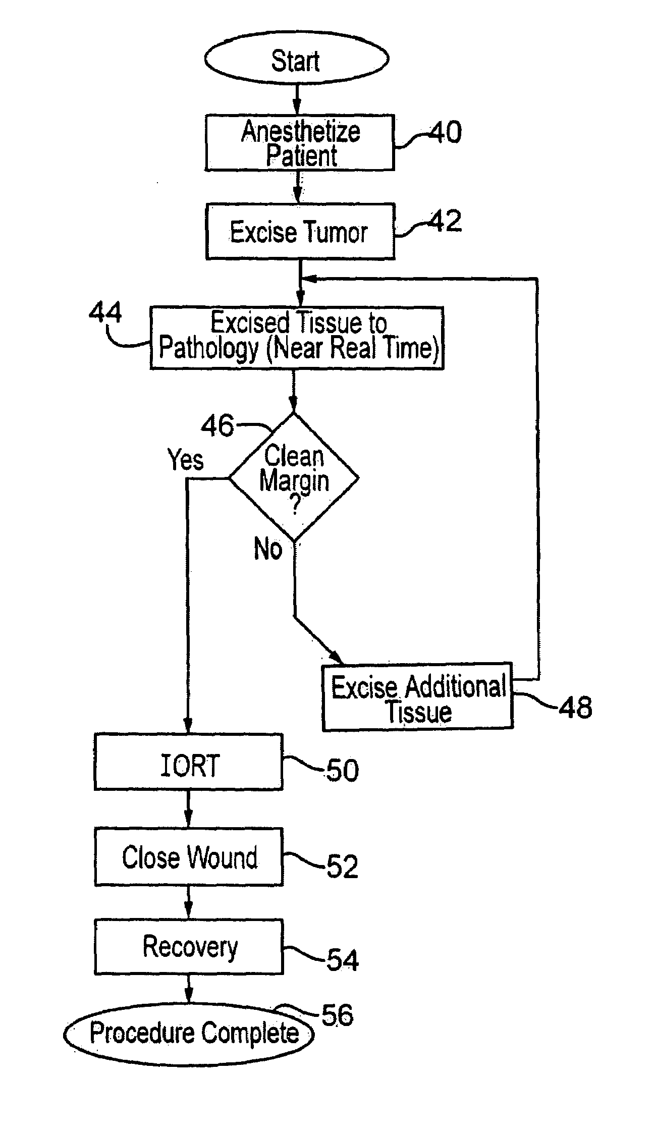
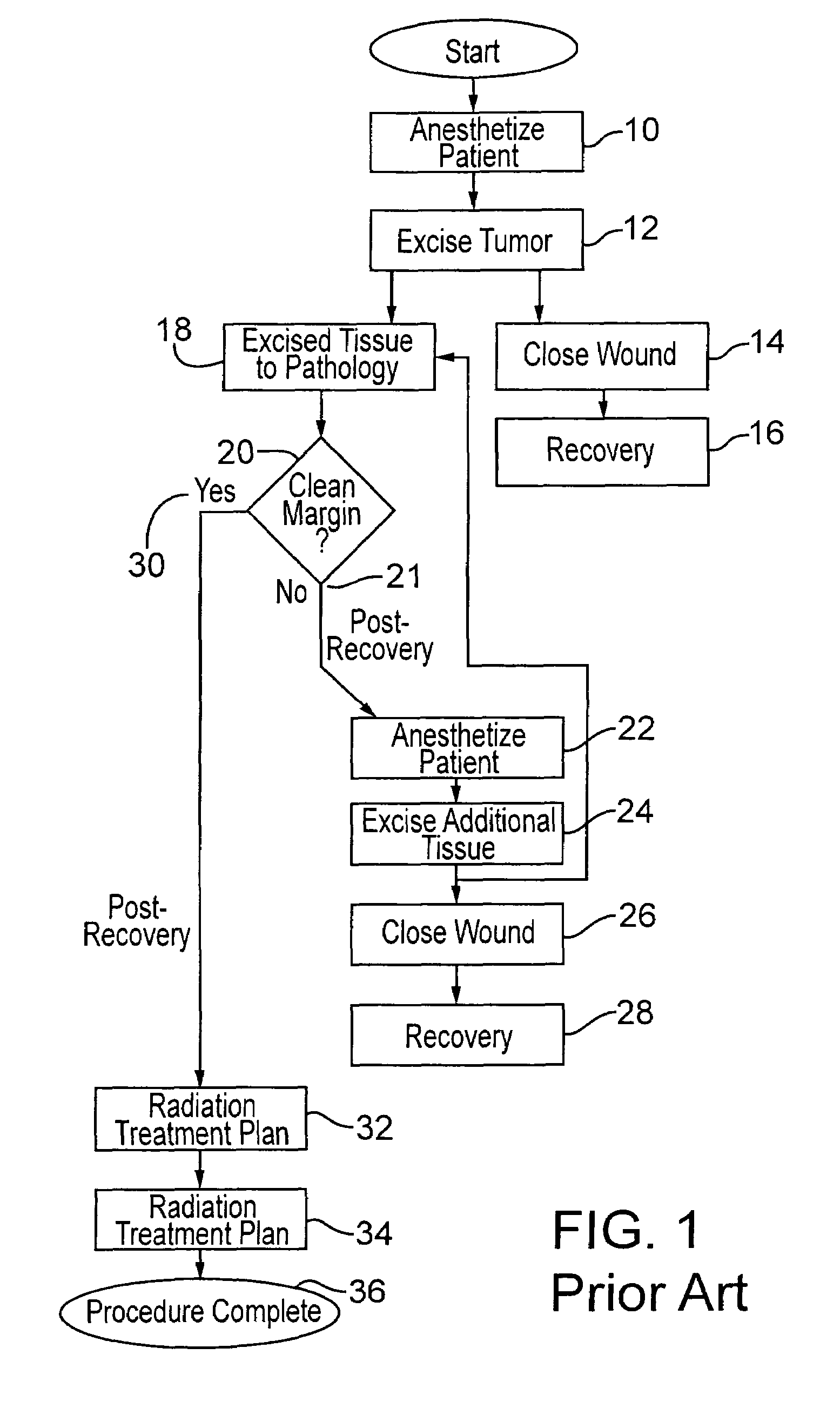
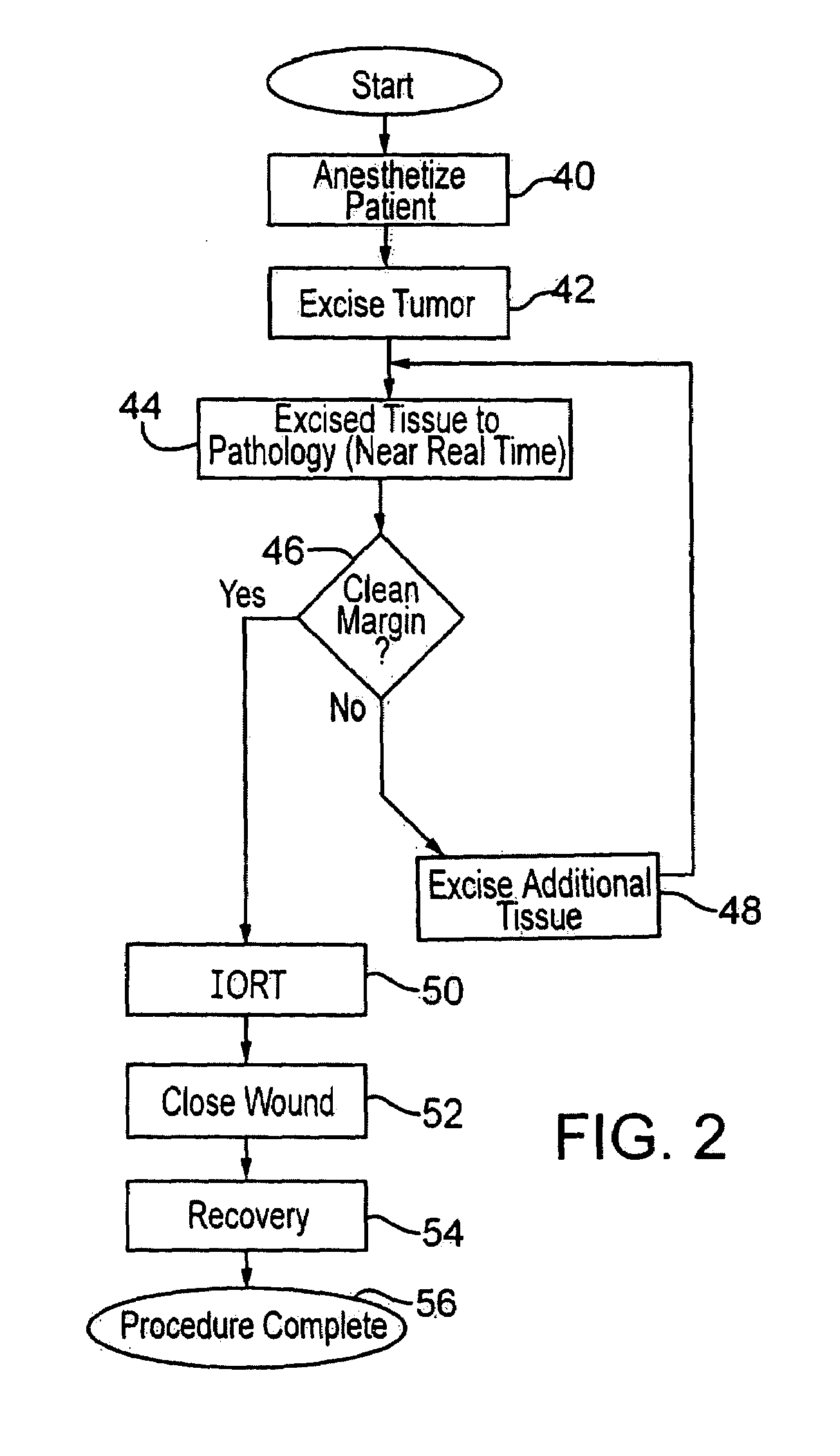
[0029]FIG. 1 shows typical current practice, prior to the present invention, for excising a tumor, particularly from the breast of a patient, and the post-operative procedure. The patient is anesthetized as indicated at 10, and the tumor is excised as indicated at 12 in the drawing. Following excision, the surgical wound is closed as noted at 14, and the patient recovers as shown in the block 16 and is sent home.

[0030]Meanwhile, the excised tissue is sent to pathology as shown at 18, and the pathology of the tissue is determined as to whether there is a clean margin, as indicated at 20. As explained above, this takes some time. Different surgeons apply different standards as to whether a margin is sufficiently clean such that radiation treatment is judged to be sufficient to remove all remaining microfoci disease which might remain. If the physician decides the pathology of the tissue does not indicate a clean margin, as at 21, then further excision is deemed to be necessary. Thus, ...


PUM


Abstract

Breast cancer patients are treated intraoperatively with radiation shortly after excision of a tumor. Pathology of the tissue is determined with a nearly instantaneous method, further excision is performed if needed, and the patient, still anesthetized and preferably unmoved, is then treated with radiation therapy. In a preferred embodiment an applicator is inserted into the excision cavity, the cavity is three-dimensionally mapped using radiation sources and a sensor, a radiation treatment plan is developed using a radiation prescription and the determined shape and location of the cavity, and the treatment plan is executed, all while the patient remains under anesthesia.

Description

BACKGROUND OF THE INVENTION[0001]This invention concerns the treatment of breast cancer or otherwise-sited cancer, and especially an efficient procedure for radiation treatment following surgical tumor excision, including pathology of the excised tissue, without waking or moving the patient or using external imaging techniques.[0002]In treating cancer of the breast, as well as cancer found in other areas of the human body, with the patient under anesthesia, the tumor is surgically excised (with some surrounding tissue) and then typically, the surgical wound is closed, the patient is sent home pending determination of pathology of the excised tumor margin, a radiation treatment plan is developed, and the patient in a series of later visits is subjected to radiation treatment in the volume of tissue surrounding the excised tumor. This can often involve re-opening of the surgical cavity for insertion of an applicator for use with ionizing radiation sources, i.e. radioactive isotopes. T...

Claims


Application Information

Patent Timeline
Patent Type & Authority Patents(United States)
IPC IPC(8): A61N5/00A61N5/10
CPCA61N5/1015A61N5/103A61N5/1048A61N2005/1018A61N5/00
Inventor LOVOI, PAUL A.
Owner XOFT INC
Why Eureka
Try Eureka