

Fluorescence electronic endoscopic system
- Summary
- Abstract
- Description
- Claims
- Application Information
AI Technical Summary
Benefits of technology
Problems solved by technology
Method used
Image

Examples
experiment i
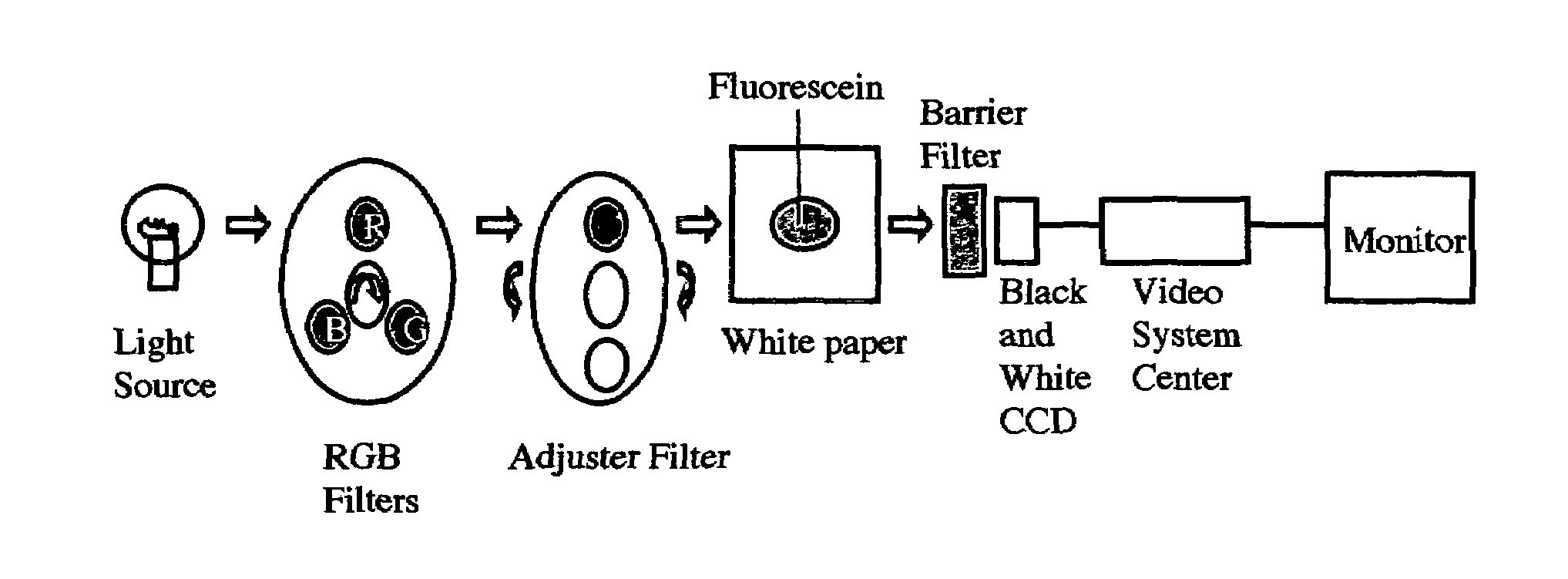
Mechanism of FEES
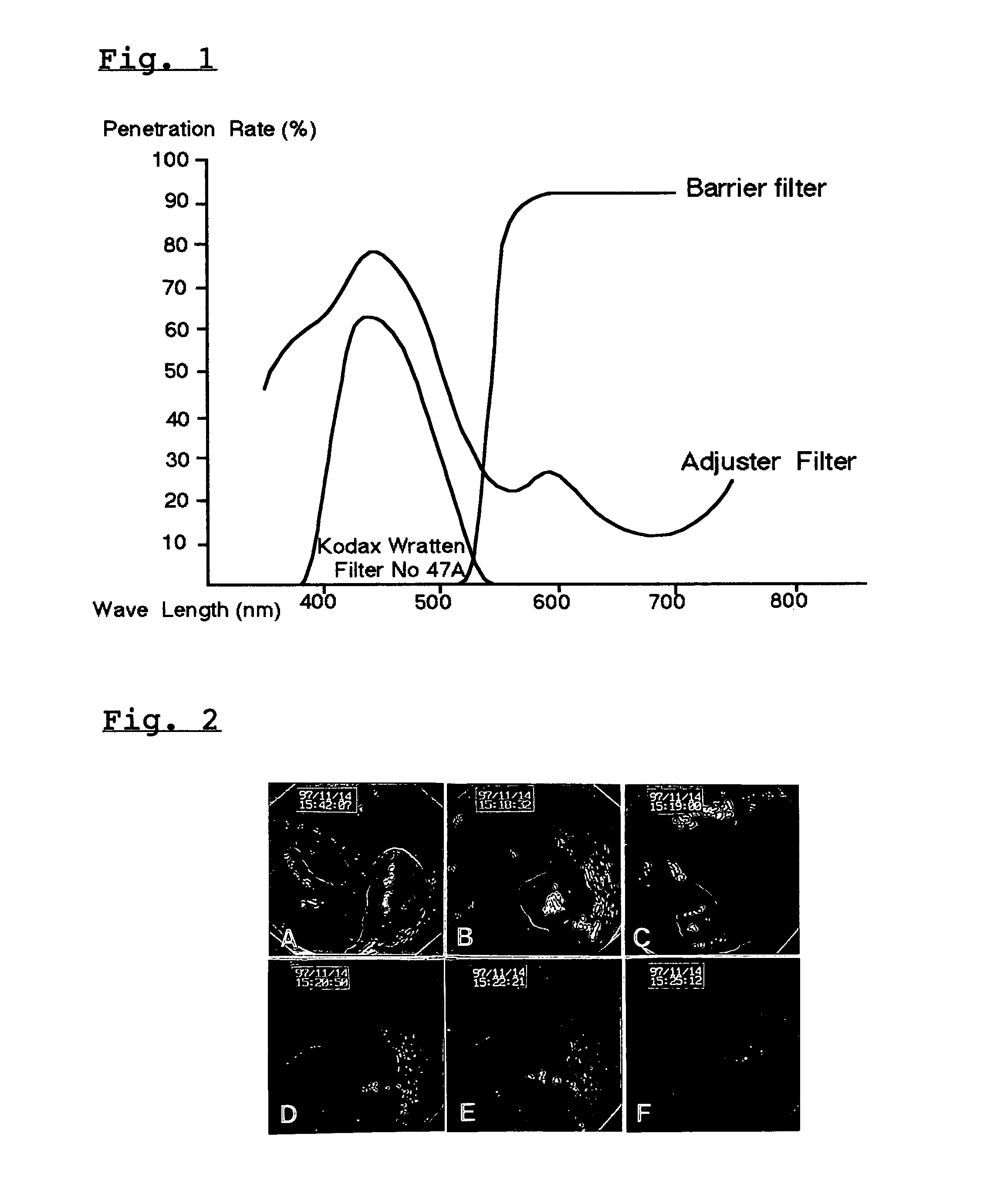
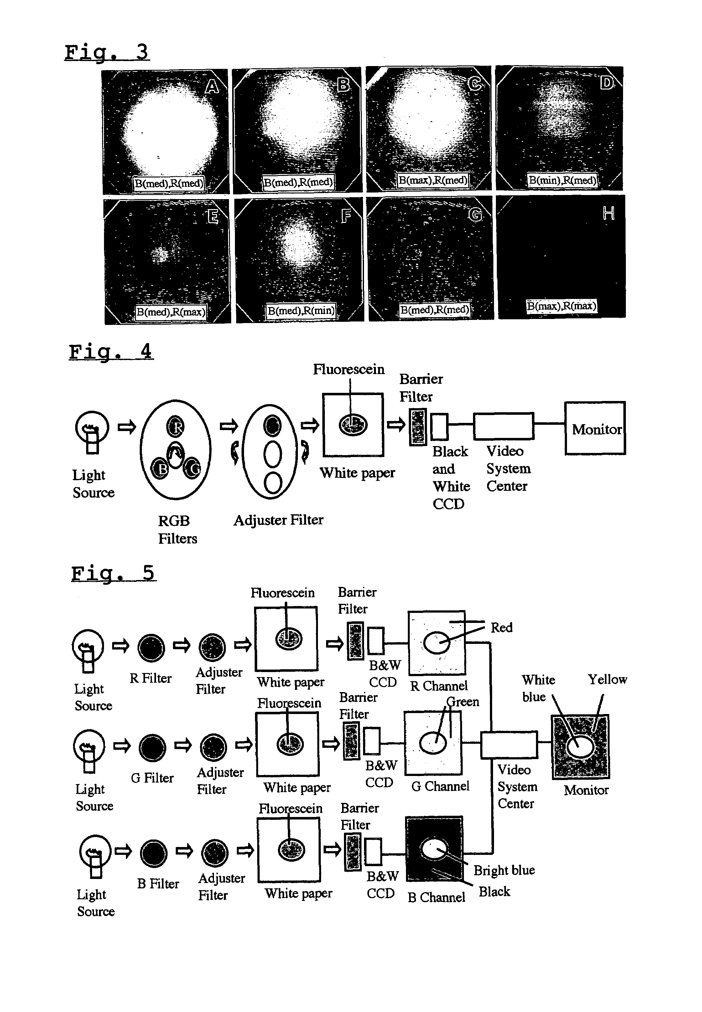
[0026]When commonly observed without a barrier filter, the stains of fluorescein sodium on white paper appeared yellowish, un-fluorescing (FIG. 3A). But, with only a barrier filter attached on the objective lens, fluorescein stains shined white fluorescence which became strongest when maximized and almost faded when the electronic signal of the blue channel was minimized. White paper appeared yellow (FIGS. 3 B, C and D). Strengthening the red channel made white paper look orange (FIG. 3E). On the other hand, weakening the red channel made white paper look green (FIG. 3F). However, strengthening and weakening the red channel did not influence the strength of white fluorescence arising on fluorescein stains (FIGS. 3E and F). With an adjuster filter in front of the light source and a barrier filter on the objective lens, white fluorescence of the fluorescein stain became stronger and its contour was clearer (FIG. 3G). White paper looked yellow but dimmer compared to wi...
experiment ii
Fluorescence Electronic Endoscopy by Fluorescein Sodium on a Rectal Polyp
[0032]Observed by white light endoscopy, the polyp and rectal wall looked reddish (FIG. 2A). With the barrier filter on the objective lens, the rectal wall looked yellow (FIG. 2B). After injection of fluorescein sodium and insertion of the adjuster filter in front of the light source, white fluorescence first appeared on the polyp several seconds later (FIG. 2C). In about 30 seconds or more, fluorescence appeared diffusely everywhere on the colonic wall (FIG. 2D). Fluorescence gradually faded but still could be observed for over ten minutes. The images of FEE is dimmer compared to routine observation but bright enough that one could see the details of the rectal wall (FIG. 2E). When an exciter filter was set instead of an adjuster filter, the images became dark blue and the details of rectal walls could no longer be observed (FIG. 2F). Fluorescence could still be seen when we switched back from the exciter filt...
PUM
 Abstract
Description
Claims
Application Information