Tissue inhibitor of matrix metalloproteinases type-1 (TIMP-1) as a cancer marker and postoperative marker for minimal residual disease or recurrent disease in patients with a prior history of cancer

a matrix metalloproteinase and tissue inhibitor technology, applied in the field of tissue inhibitors of matrix metalloproteinases type-1 (timp-1), can solve the problems of low sensitivity, lack of compliance of current tests, and significant risk of disease death, and achieve the effect of equal clinical value and level of specificity of invention

Inactive Publication Date: 2008-05-20
RIGSHOSPITALET +1
View PDF32 Cites 1 Cited by
  • Summary
  • Abstract
  • Description
  • Claims
  • Application Information

AI Technical Summary

Benefits of technology

[0003]The test is based on the measurement of tissue inhibitor of metalloproteinases type 1 (TIMP-1), in various body fluids, including blood, plasma, serum, saliva, stool, cerebrospinal fluid and urine. TIMP-1 concentrations can be determined either as the total TIMP-1 concentration, the free TIMP-1 concentration, the concentration of complexes between TIMP-1 and Matrix Metallo-Proteinases (MMPs) and / or ratios and fractions thereof, hereafter referred to as TIMP-1 levels. According to the invention, individuals with a high likelihood of having cancer, e.g. colorectal cancer can be identified by elevated TIMP-1 levels in their body fluids, while individuals with low TIMP-1 levels are unlikely to suffer from cancer, e.g. colon cancer. Also according to the invention, individuals with a prior diagnosis of cancer and who have a high likelihood of later developing clinically manifest recurrent cancer, e.g. gastrointestinal cancer such as colon cancer, can be identified by elevated postoperative TIMP-1 levels in their body fluids, while individuals with low postoperative TIMP-1 levels are unlikely to suffer from or later develop recurrent cancer. Thus, the invention can be used to identify individuals with a high probability of having early stage, non-symptomatic cancer, e.g. colon cancer or later developing recurrent colorectal cancer. The identified individuals should be further examined and if cancer, recurrent cancer or minimal residual disease (MRD) or both recurrent cancer and MRD are found, the patients should be offered surgery, irradiation, anti-neoplastic therapy or any combination thereof, thereby increasing the chance of cure and / or long term survival of the individual.

Problems solved by technology

Following relapse, the risk of dying of the disease is significant.
Screening for colorectal cancer has been shown to improve survival, however, current tests suffer from a lack of compliance, from low sensitivity, and from the need for strict dietary restrictions.
Thus, 30-40 percent of patients who have undergone a complete resection of a colorectal malignancy will experience relapse with metastatic disease, which is usually fatal.

Method used


Image

  • Tissue inhibitor of matrix metalloproteinases type-1 (TIMP-1) as a cancer marker and postoperative marker for minimal residual disease or recurrent disease in patients with a prior history of cancer
  • Tissue inhibitor of matrix metalloproteinases type-1 (TIMP-1) as a cancer marker and postoperative marker for minimal residual disease or recurrent disease in patients with a prior history of cancer
  • Tissue inhibitor of matrix metalloproteinases type-1 (TIMP-1) as a cancer marker and postoperative marker for minimal residual disease or recurrent disease in patients with a prior history of cancer

Examples


example 1

[0113]Preparation of an ELISA to quantitate total TIMP-1 concentrations in human plasma.

[0114]This example describes the preparation and validation of an ELISA that measures total TIMP-1 levels in plasma. In addition, this example provides information on TIMP-1 levels in different plasma preparations as well as in healthy blood donors of both sexes.

Materials and Methods

Blood Donors

[0115]Blood samples were initially obtained from 94 apparently healthy volunteer blood donors, comprising 51 males aged 19 to 59 years (median: 41 years) and 43 females aged 20 to 64 years (median: 36 years). In a subsequent collection, 100 donor samples were obtained, comprising 56 males aged 19 to 59 years (median 42: years) and 44 females aged 20 to 60 years (median: 36.5 years). Informed consent was obtained from all donors, and permission was obtained from the local Ethical Committees.

Blood Collections and Plasma Separation

[0116]Peripheral blood was drawn with minimal stasis (if necessary a maximum of...

example 2

[0136]Preparation of an ELISA to quantitate TIMP-1:MMP-9 complexes in plasma.

[0137]The following example describes an assay to determine the concentration of TIMP-1:MMP-9 complexes in body fluids. The assay is used with plasma samples of healthy blood donors in order to establish normal ranges of this complex (Holten Andersen et. al., 1999).

Materials and Methods

TIMP-1: MMP-9 Complex ELISA

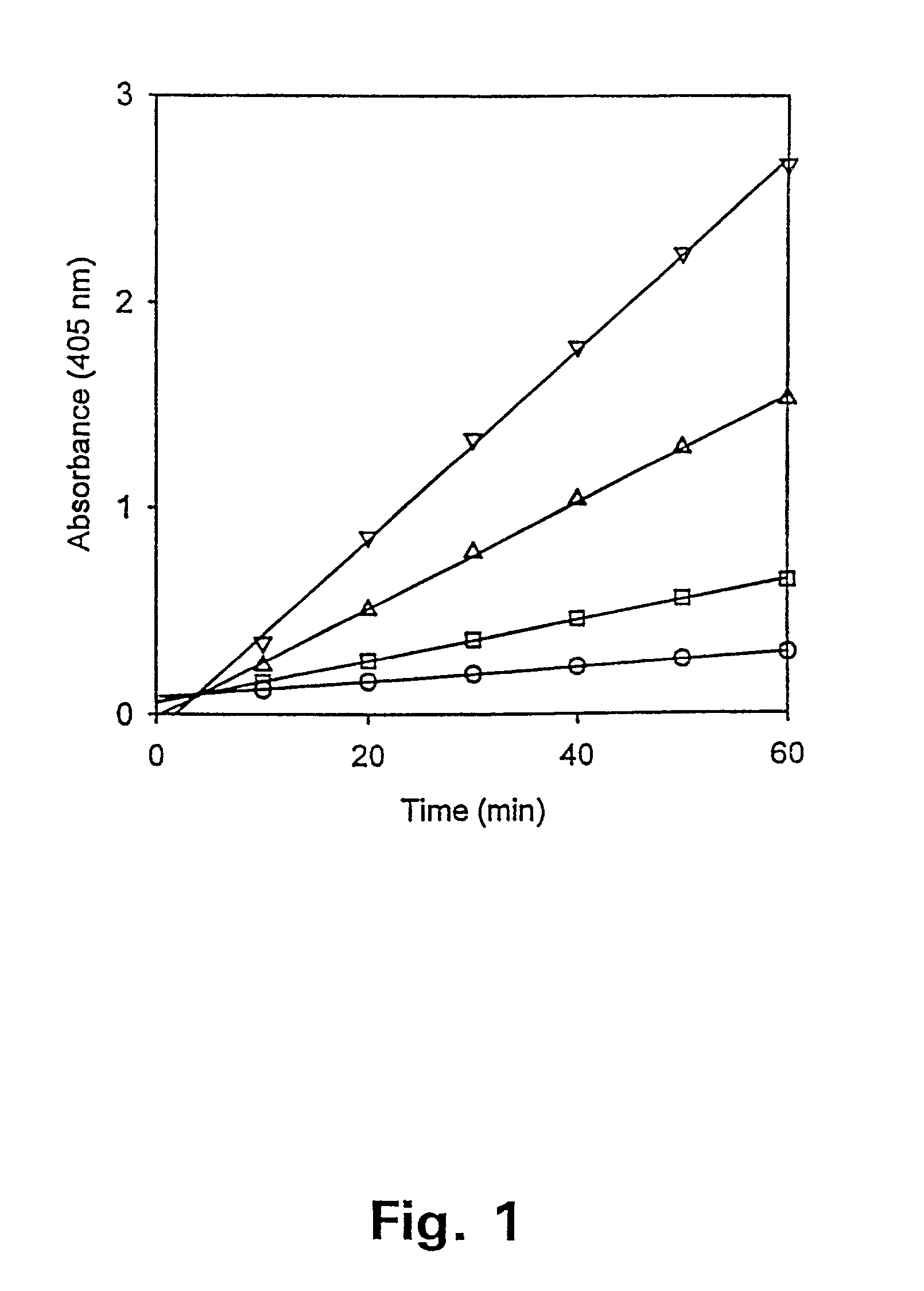
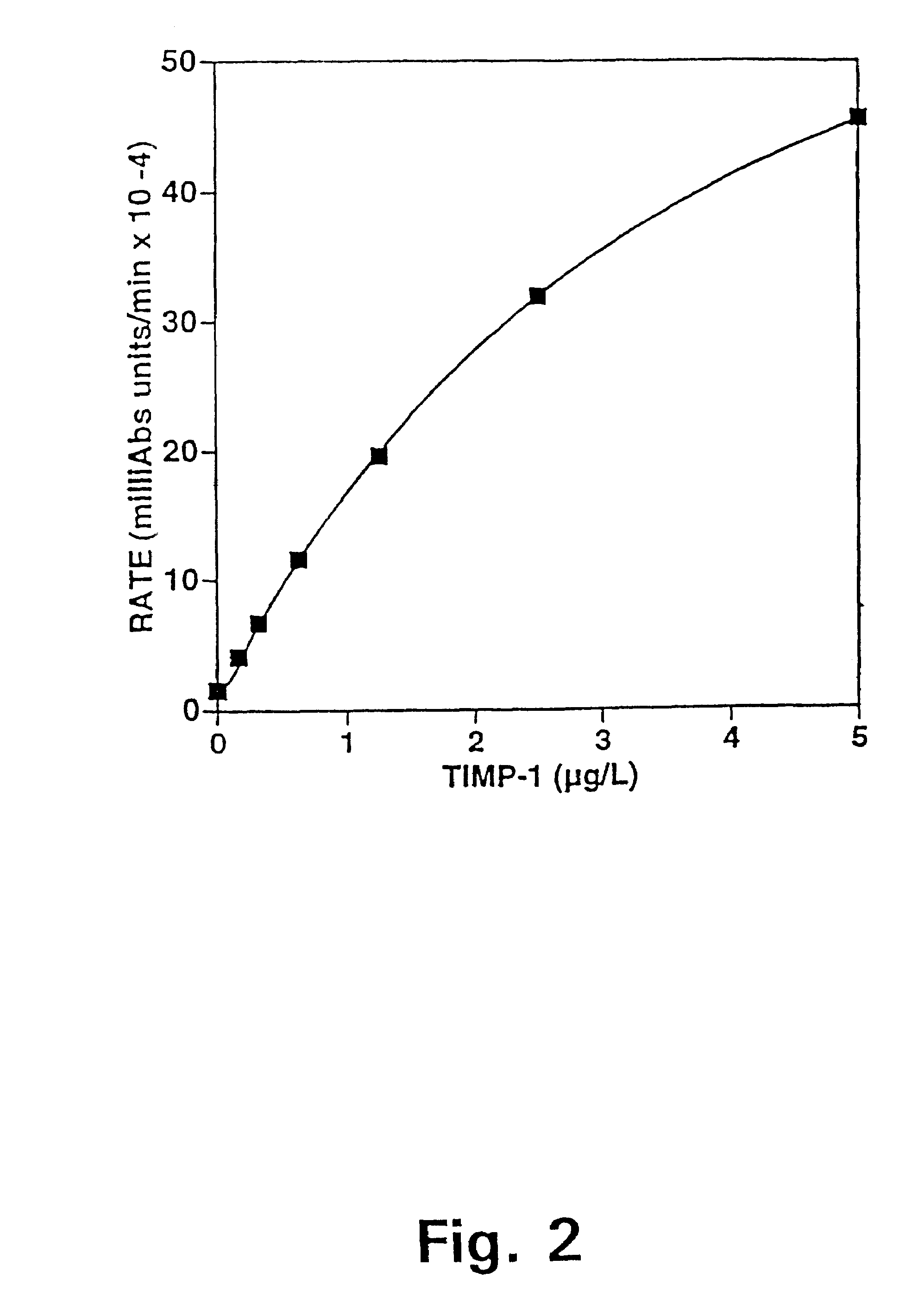
[0138]A sensitive and specific sandwich ELISA was prepared using the above-described TIMP-1 antibody, MAC-15, and a rabbit MMP-9 polyclonal antibody developed in the Hematological Department, Rigshospitalet, Denmark (Kjeldsen et. al., 1992). The MMP-9 antibody was used for antigen capture and MAC-15was used for antigen detection. A rabbit anti-mouse-Ig / alkaline phosphatase conjugate (DAKO®, Glostrup, Denmark) enabled a kinetic rate assay (FIG. 8). The latter conjugate was supplied preabsorbed against human IgG, thus eliminating cross-reactivity with IgG in the plasma samples. The MMP-9 antibody capt...

example 3

[0146]Preparation of an ELISA to quantitate free TIMP-1 levels in plasma.

[0147]The following example describes an assay that determines the concentration of free TIMP-1 levels in body fluids. The assay is applied to plasma samples of healthy blood donors in order to establish normal ranges of free TIMP-1.

Materials and Methods

FREE TIMP-1 ELISA

[0148]A sensitive and specific sandwich immunoassay was prepared, using a TIMP-1 monoclonal IgG1 antibody (MAC-19) developed at the Strangeways Laboratories, England (Cooksley et. al., 1990) and a sheep polyclonal anti-TIMP-1 antibody. The sheep polyclonal anti-TIMP-1 antibody was used for antigen capture and the murine monoclonal MAC-19 was used for antigen detection. A rabbit anti-mouse-Ig / alkaline phosphatase conjugate was the secondary detection reagent (FIG. 9). The latter conjugate was supplied preabsorbed against human IgG, thus eliminating cross-reactivity with IgG in the plasma samples. The MAC-19 monoclonal antibody is completely speci...


PUM

PropertyMeasurementUnit
timeaaaaaaaaaa
timeaaaaaaaaaa
timeaaaaaaaaaa

Abstract

The present invention describes a method for determining whether an individual is suffering from cancer by determining a parameter representing the TIMP-1 concentration in body fluid samples from the individual.The present invention furthermore describes a method for determining whether an individual is suffering from minimal residual disease or recurrent cancer after being treated for the primary cancer by determining a parameter representing the post-operative TIMP-1 concentration in body fluid samples from the individual. In addition, the invention describes the additive effect of combined post-operative measurements of plasma TIMP-1 and serum CEA.

Description

CROSS-REFERENCE TO RELATED APPLICATIONS[0001]This application is a continuation-in-part of application Ser. No. 09 / 546,573, filed Apr. 10, 2000 (the “'573 Application”), now U.S. Pat. No. 7,108,983, issued on Sep. 19, 2006, which claims priority to Denmark Patent Application No. DK 1999 00476 filed Apr. 9, 1999. The entire contents of the '573 Application are incorporated herein by reference to the extent they are consistent with this application and invention.BRIEF DESCRIPTION OF THE INVENTION[0002]The present invention relates to a test to be used to screen large populations for the occurrence of cancer. Furthermore it relates to a method for monitoring colorectal cancer patients for the persistency of minimal residual disease (MRD) or for the recurrence of their cancer disease. The method is based on the measurement of tissue inhibitor of metalloproteinases 1 (TIMP-1) in body fluids either alone or in combination with other tumor markers, e.g. Carcino Embryonic Antigen (CEA). The...

Claims


Application Information

Patent Timeline
Patent Type & Authority Patents(United States)
IPC IPC(8): G01N33/53G01N33/574
CPCG01N33/57415G01N33/57419G01N33/57484G01N2333/8146G01N2333/96494
Inventor HOLTEN-ANDERSEN, MADS NIKOLAJCHRISTENSEN, IB JARLEBRUNNER, NILSNIELSEN, HANS JORGEN
Owner RIGSHOSPITALET
Why Eureka
Try Eureka