Prion protein binding materials and methods of use

a technology of protein binding and materials, applied in the field of protein binding, can solve the problems of inability to identify infected animals using conventional serologic tests, no antibodies specific for tscs have been identified, and no known nucleic acid sequence can be used in polymerase chain reaction-based diagnostic methods

Active Publication Date: 2008-07-01
PATHOGEN REMOVAL & DIAGNOSTIC TECH +1
View PDF18 Cites 38 Cited by
  • Summary
  • Abstract
  • Description
  • Claims
  • Application Information

AI Technical Summary

Benefits of technology

[0011]Materials that bind to prion proteins and methods for using the prion protein binding materials (hereinafter “binding materials”) are provided. In some embodiments, the binding materials are polymer particles, preferably chromatographic resins, that bind with selectivity and specificity to prion analytes. In other embodiments, the binding materials are inorganic materials that bind with selectivity and specificity to prion analytes. The binding materials are capable of binding to one or more forms of prion protein including cellular prion protein (PrPc), infectious prion protein (PrPsc), and recombinant prion protein (PrPr). Prions from various species, including humans and hamsters, are bound by the binding materials. Compositions containing the binding materials on a support such as a chromatography column are also provided.

Problems solved by technology

Consequently, no antibodies specific for TSCs have been currently identified.
Moreover, the lack of a known nucleic acid sequence precludes the use of polymerase chain reaction-based diagnostic methods.
Thus, no conventional serologic test can be used to identify infected animals.
Other factors elevating concern about the risk of transmission by transfusion include the unknown, but presumably high, numbers of people exposed to BSE and lack of a preclinical diagnostic test for vCJD.
Unfortunately, TSE infectivity is remarkably resistant to chemical and physical methods of inactivation, and a selective method of inactivation is elusive.
Currently, no material has been fully characterized or found to be able to bind to prion from a wide variety of media.
To date, human TSE diseases are 100% fatal.
Unfortunately, even though a number of compounds including amphotericins, sulphated polyanions, Congo Red dye and anthracycline antibiotics have been reported as prospective therapeutic agents, all have demonstrated only modest potential to impede prion propagation, and none have been shown to have any effect on the removal of pre-existing prions from an infected host in a controlled clinical study.

Method used


Image

  • Prion protein binding materials and methods of use
  • Prion protein binding materials and methods of use
  • Prion protein binding materials and methods of use

Examples


example 1

Identification of Prion-binding Materials

[0084]Eighty polymer or inorganic particles were tested by using a prion binding on-beads test by a NBT / BCIP chromagen (nitroblue tetrazolium / 5-bromo-4-chloro-3-indolyl-phosphate-p-toluidine salt), as described below, using normal hamster brain homogenate. The binding results are provided in Table 1 wherein “−” means no binding and “+” means positive binding. The more “+” in a particle rating, the stronger the binding observed. Twelve particles that had at least “++” were evaluated further. Table 2 summarizes the twelve particles in their ability to bind to normal hamster prion. A higher score indicates an increased amount of prion binding.

[0085]

TABLE 1Screening Polymeric Material or Inorganic Particles for Prion Protein BindingReference No.NameManufacturerBinding Results 1Toyopearl ™ Amino-650MTosoh Bioscience+++(Montgomeryville, PA) 1′Acetylated Amino-650MAcetylation done by North−Carolina State University(NCSU) using Toyopearl ™Amino 650M ...

example 2

Identification of Prion-Binding Materials

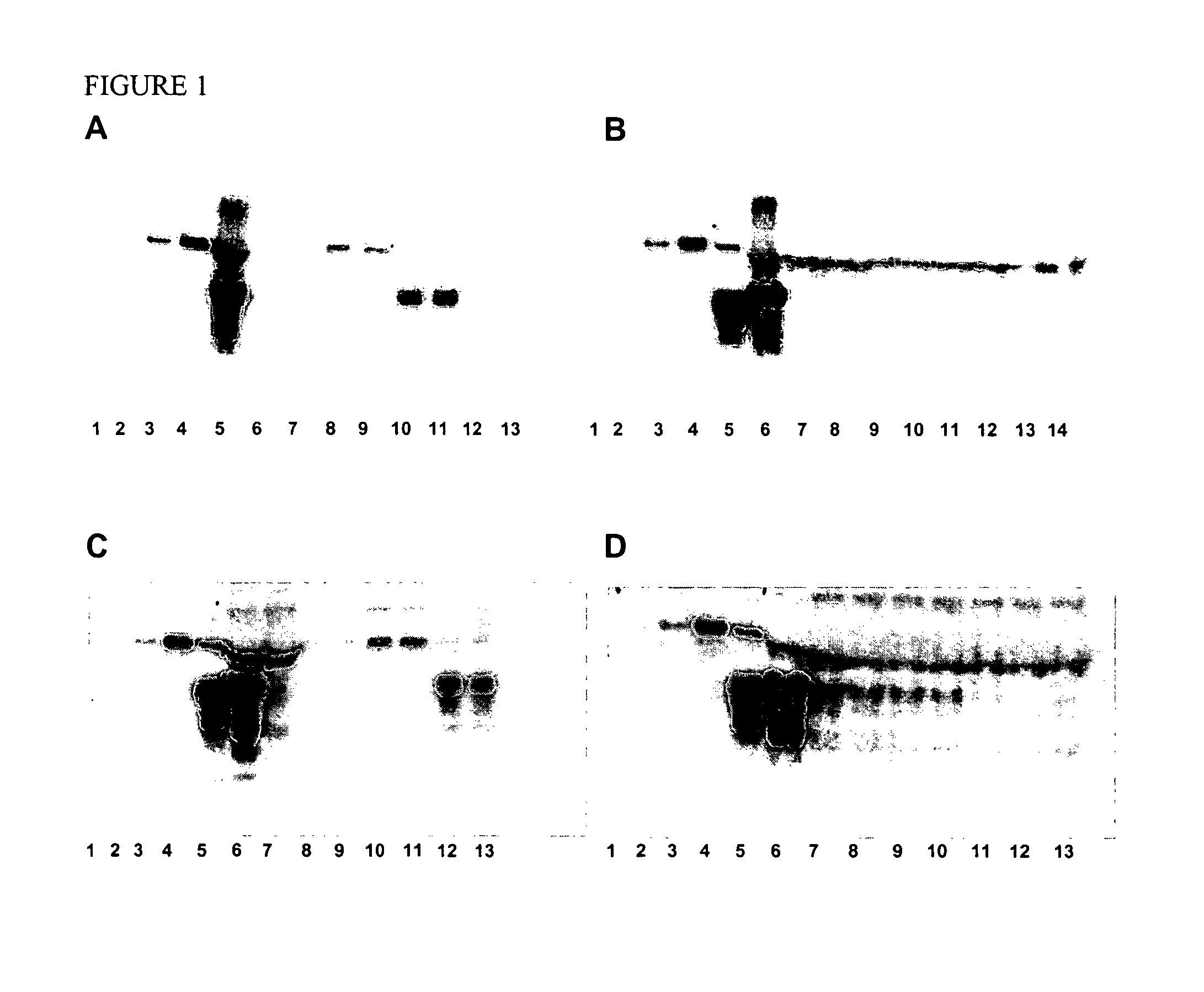
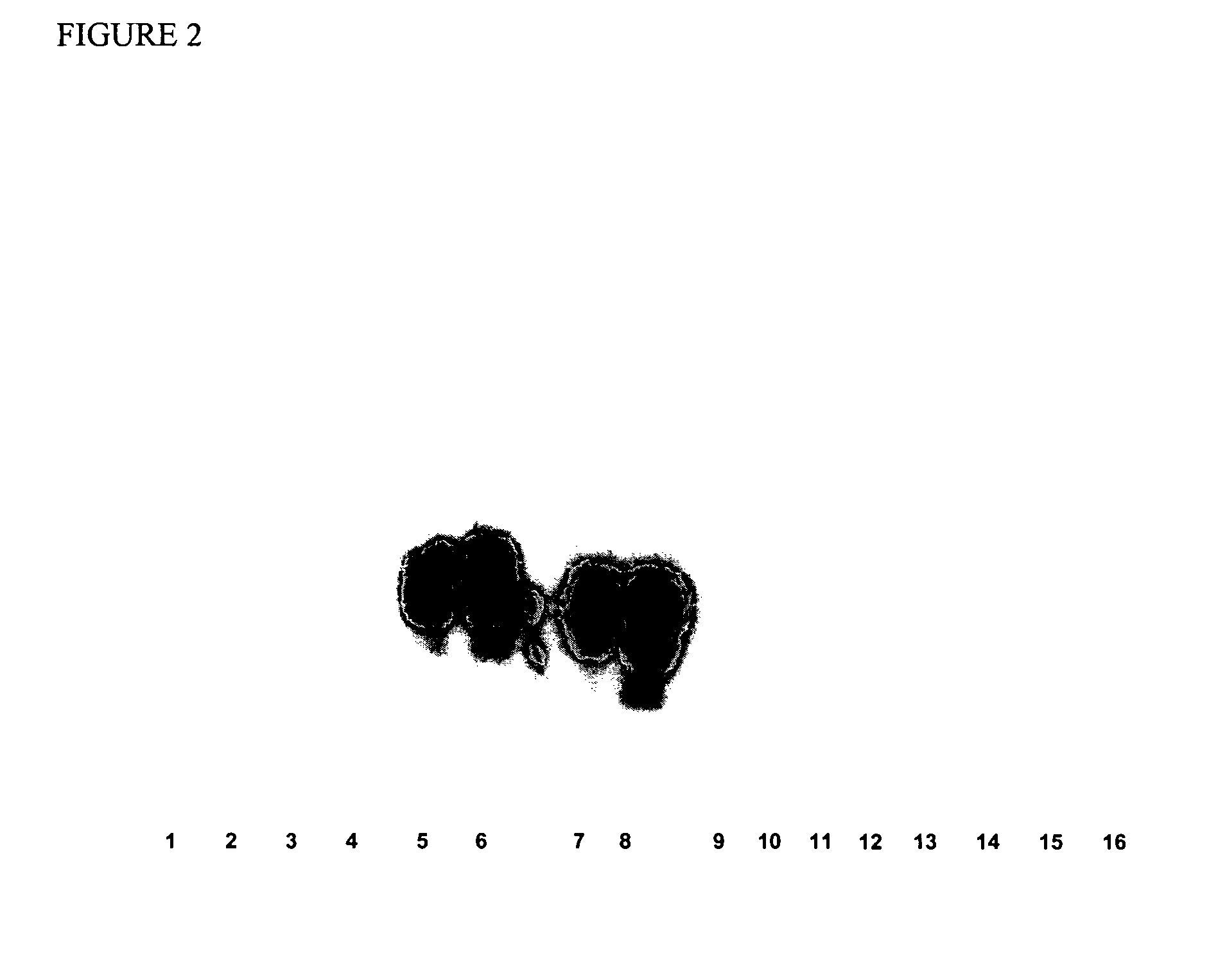
[0091]Identification of prion-binding materials was performed using hamster brain homogenate in batch format, using two different detection systems. In the first, the amount of prion bound to a material was detected by staining the beads after incubation with the target material. The second method detected the amount of prion present in the unbound fraction contained in flow-through and wash samples using SDS-PAGE and western blots. A detailed description of each methodology is described below.

[0092]As shown in Table 3 below, the Toyopearl™ Amino-650M, TSK-GEL™ Amino 750C and TSK-GEL™ Phenyl-5PW provided the most specific binding of hamster brain PrPc.

[0093]

TABLE 3Results of Secondary ScreeningBindingBinding evaluatedevaluated byby on-bead testswestern blot withPolymer Particleswith NBT / BCIPECL-plusToyopearl ™ Amino-650M++++++TSK-GEL ™ Amino 750C++++++TSK-GEL ™ Phenyl-5PW+++++++Toyopearl ™ Butyl-650C+++Toyopearl ™ Phenyl-650C+++TSK-GEL ™ DEAE...

example 3

Determination of Prion-binding Specificity

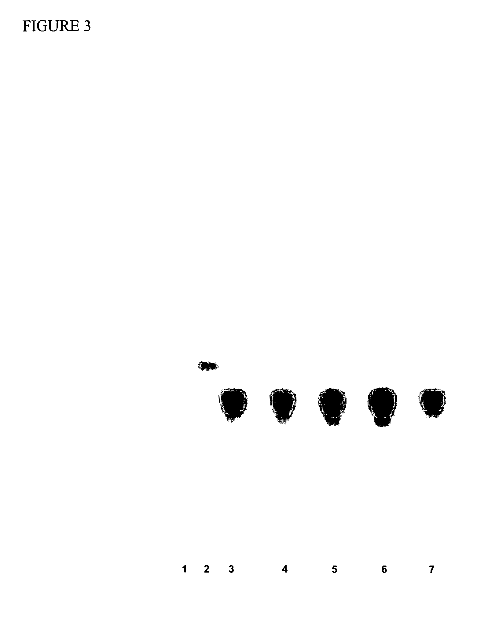
[0100]Generally, wetted beads composed of different binding materials were quantitatively placed into individual disposable columns. The columns contained frits small enough to retain the beads but large enough to permit flow-through of the challenge solutions. The challenge solutions were prion-containing brain homogenates in Sarkosyl (Sigma) spiked into red blood cell concentrate comixtures. More specifically, the challenge solutions contained TSE-infectious human brain homogenates, infectious hamster brain homogenates, noninfectious human brain homogenates, or noninfectious hamster brain homogenates. The challenge solutions were allowed to pass through the target binding material for a defined period of time, while the flow-through was being collected. Beads were then rinsed and quantitatively transferred from their columns into collection vials from which known quantities were removed for subsequent processing to determine specific prion...


PUM

PropertyMeasurementUnit
mean particle sizeaaaaaaaaaa
molecular weightaaaaaaaaaa
sizeaaaaaaaaaa

Abstract

Prion protein binding materials and methods for using the binding materials to detect or remove a prion protein from a sample, such as a biological fluid or an environmental sample. The binding materials are capable of binding to one or more forms of prion protein including cellular prion protein (PrPc), infectious prion protein (PrPsc), recombinant prion protein (PrPr), and proteinase resistant prion protein (PrPres). Prions from various species, including humans and hamsters, are bound by the binding materials.

Description

CROSS-REFERENCE TO RELATED APPLICATIONS[0001]This application claims benefit of U.S. Provisional Patent Application Ser. No. 60 / 460,474 filed Apr. 4, 2003.FIELD OF THE INVENTION[0002]This invention relates to the field of protein binding and more particularly relates to materials that bind to prion proteins and methods of using the prion protein binding materials to detect or remove prions from biological samples.BACKGROUND OF THE INVENTION[0003]Native or cellular prion protein “PrPc” is widely distributed throughout the mammalia and has a particularly well-conserved amino acid sequence and protein structure. Infectious prions are thought to be composed of a modified form of the normal cellular (PrPc) prion protein and are called “PrPsc”. Prions have some properties in common with other infectious pathogens, but do not appear to contain nucleic acid. Instead, it is proposed that a post-translational conformational change is involved in the conversion of non-infectious PrPc into infe...

Claims


Application Information

Patent Timeline
Patent Type & Authority Patents(United States)
IPC IPC(8): G01N33/53C12NG01N33/68
CPCG01N33/6896
Inventor CARBONELL, RUBEN G.SHEN, HONGLUEGURGEL, PATRICK V.WILTSHIRE-LYERLY, VITEROSHAMMOND, DAVID J.BURTON, STEVEN J.
Owner PATHOGEN REMOVAL & DIAGNOSTIC TECH
Why Eureka
Try Eureka