Method and apparatus for treating pelvic organ prolapse

a pelvic organ and prolapse technology, applied in the field of pelvic organ prolapse treatment methods and equipment, can solve the problems of increasing abdominal pressure, increasing risk for patients, and increasing problems, and achieves the effects of less risk for patients, convenient operation, and simple operation

Active Publication Date: 2009-03-10
ASTORA WOMENS HEALTH
View PDF210 Cites 264 Cited by
  • Summary
  • Abstract
  • Description
  • Claims
  • Application Information

AI Technical Summary

Benefits of technology

[0029]The present invention is directed to a method and apparatus for treating pelvic organ prolapse, and a kit containing elements for practicing the same. The present invention includes a support member less susceptible to deformation, relative to the prior art, following implantation and a means for repositioning and adjusting the support member which does not subject the sling to deformation pressures. The method of treatment is one that allows the operator to know the location of the instruments, as final passage of the needle is aided by the operator's use of his finger, making the method less risky for the patient. The apparatus and method is convenient for the operator, in that the apparatus is relatively simple to operate and contained within the described kit. The sling portion is relatively extensible compared to the prior art. The needle is of a small diameter to reduce the risk of trauma.
[0036]In another embodiment, the apparatus is substantially one tape, in which the support portion is a wider center section, relative to the two end portions, in which the support portion and the end portions are substantially one tape. Such an embodiment would allow for easier and more secure suture attachment.
[0037]In another embodiment, the support portion is of a different material in order to provide for better suture retention.

Problems solved by technology

Female genital prolapse has long plagued women.
With the increasing age of the U.S. population, these problems will likely assume additional importance.
Increases in abdominal pressure, failure of the muscles to keep the pelvic floor closed, and damage to the ligaments and fascia all contribute to the development of prolapse.
In addition, if a woman has a hysterectomy, the vaginal angle may be altered, causing increased pressure at a more acute angle, accelerating the prolapse.
Further, loss of connective tissue strength might be associated with damage at childbirth, deterioration with age, poor collagen repair mechanisms, and poor nutrition.
The common clinical symptoms of vaginal prolapse are related to the fact that, following hysterectomy, the vagina is inappropriately serving the role of a structural layer between intra-abdominal pressure and atmospheric pressure.
This pressure differential puts tension on the supporting structures of the vagina, causing a “dragging feeling” where the tissues connect to the pelvic wall or a sacral backache due to traction on the uterosacral ligaments.
Exposure of the moist vaginal walls leads to a feeling of perineal wetness and can lead to ulceration of the exposed vaginal wall.
Vaginal prolapse may also result in loss of urethral support due to displacement of the normal structural relationship, resulting in stress urinary incontinence.
Certain disruptions of the normal structural relationships can result in urinary retention, as well.
Stretching of the bladder base is associated with vaginal prolapse and can result in complaints of increased urinary urgency and frequency.
Anterior vaginal wall prolapse causes the vaginal wall to fail to hold the bladder in place.
There may also be a transverse defect, causing cystecele across the vagina.
They may occur because of failure to reapproximate the superior aspects of the pubocervical fascia and the rectovaginal fascia at the time of surgery.
Mid-vaginal and high rectoceles may result from loss of lateral supports or defects in the rectovaginal septum.
As noted, vaginal prolapse and the concomitant anterior cystocele can lead to discomfort, urinary incontinence, and incomplete emptying of the bladder.
Posterior vaginal prolapse may additionally cause defecatory problems, such as tenesmus and constipation.
Vaginal pessaries are the primary type of nonsurgical treatment, but there can be complications due to vaginal wall ulceration.
In addition, it is often required to correct multiple pelvic floor abnormalities simultaneously, further increasing surgical time.
This procedure requires specialized skills and has the further disadvantage of tending to place the vagina in an artificial anatomical position.
Interestingly, relief of pelvic organ prolapse often results in incontinence in the patient.
Although serious complications associated with sling procedures are infrequent, they do occur.
Complications associated with the TVT procedure and other known sling procedures include injury to blood vessels of the pelvic sidewall and abdominal wall, hematomas, urinary retention, and bladder and bowel injury due to passage of large needles.
One serious disadvantage of the TVT procedure, particularly for surgeons unfamiliar with the surgical method, is the lack of information concerning the precise location of the needle tip relative to adjacent pelvic anatomy.
If the needle tip is allowed to accidentally pass across the surface of any blood vessel, lymphatic duct, nerve, nerve bundle or organ, serious complications can arise.
Additional problems are associated with the TVT and other sling procedures.
However, the smooth surface of the needles, which facilitates insertion through the tissues, prevents secure attachment of the forceps onto the needles, causing slippage or detachment of the forceps during the withdrawal procedure.
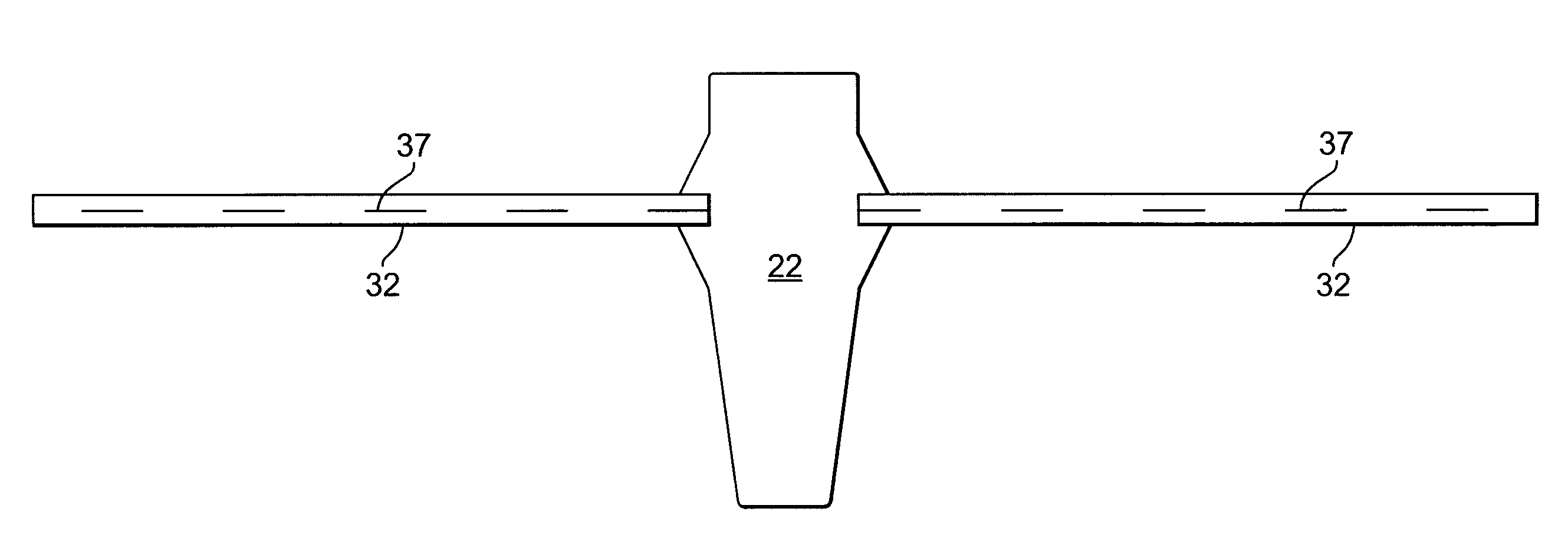
Improper placement of the TVT mesh is also particularly troublesome.
If the mesh is too loosely associated with its intended physiological environment, the mesh may be ineffective in supporting the urethra and treating incontinence.
Several complications can arise from a mesh that is too tightly placed including retention, sling erosion and other damage to surrounding tissue such as the urethra and vagina.
Surgeons may exacerbate these problems by improperly attempting to adjust the tension of a sling.
If insufficient adjustment force is applied, the sling will simply exhibit a memory property and return to its original, unacceptable position.
As a result, surgeons are tempted to use a great deal of force in order to loosen a sling that is perceived to be too tightly associated with its intended physiological environment.
Excessive deformation may result in a lack of efficacy or, even worse, the edges of the mesh may curl up and present a relatively sharp, frayed surface.
In this curled or deformed state, the edges of the TVT mesh present sharp surfaces that can readily abrade or otherwise damage adjacent tissue such as the urethra, bladder or vagina.
The problems associated with the TVT mesh device are commonly seen in other similar sling or synthetic implant devices.
This tube passes through two separate regions of the patient's body with the attendant risk of cross-contamination.
The outer diameter is also relatively large (about 0.25 inches) with the attendant risk of tissue damage due to such large diameter.
This tape is not believed to be optimally sized and shaped to afford concomitant procedures such as enterocele, cystocele, and or rectocele repairs.
The tape is also largely inextensible.
Such inextensibility is believed to be associated with higher risk of tissue erosion and failure.

Method used


Image

  • Method and apparatus for treating pelvic organ prolapse
  • Method and apparatus for treating pelvic organ prolapse
  • Method and apparatus for treating pelvic organ prolapse

Examples


Embodiment Construction

[0057]Referring now to the drawings, wherein like reference numerals designate identical or corresponding parts throughout the several views. The following description is meant to be illustrative only and not limiting. Other embodiments of this invention will be apparent to those of ordinary skill in the art in view of this description.

[0058]Two tissue pathways are established between the external perirectal region and the region of the ischial spine of the patient. These pathways are made by making incisions in the rectal area and the vaginal apex and passing a needle through the rectal area incision toward the vaginal incision. Referring now to the drawings, wherein like reference numerals designate identical or corresponding parts throughout the several views, FIG. 1 shows a needle 14 and handle 10 suitable for use in the present invention. The handle 10 can be any suitable handle known in the art. U.S. Pat. No. 6,652,450, hereby incorporated by reference in its entirety, disclos...


PUM


Abstract

A method of treating pelvic organ prolapse is provided. The method generally includes the steps of establishing a first pathway between the external perirectal region of the patient to the region of the ischial spine in tissue on one side of the prolapsed organ, followed by establishing a second pathway in tissue on the contralateral side of the prolapsed organ. A support member, which includes a central support portion and two end portions, is positioned in a position to reposition said prolapsed organ in said organ's anatomically correct location. The end portions of the support member are introduced through the respective tissue pathways, followed by adjustment of the end portions so that the support member is located in a therapeutic relationship to the prolapsed organ that is to be supported. An apparatus and kit for said treatment is further provided.

Description

BACKGROUND OF THE INVENTION[0001]1. Field of the Invention[0002]Urogenital Surgery[0003]2. Description of the Related Art[0004]Female genital prolapse has long plagued women. It is estimated by the U.S. National Center for Health Statistics that 247,000 operations for genital prolapse were performed in 1998. With the increasing age of the U.S. population, these problems will likely assume additional importance.[0005]Vaginal prolapse develops when intra-abdominal pressure pushes the vagina outside the body. In a normal situation, the levator ani muscles close the pelvic floor. This results in little force being applied to the fascia and ligaments that support the genital organs. Increases in abdominal pressure, failure of the muscles to keep the pelvic floor closed, and damage to the ligaments and fascia all contribute to the development of prolapse. In addition, if a woman has a hysterectomy, the vaginal angle may be altered, causing increased pressure at a more acute angle, acceler...

Claims


Application Information

Patent Timeline
Patent Type & Authority Patents(United States)
IPC IPC(8): A61F2/00A61B17/00A61B17/04A61B17/06
CPCA61B17/06109A61F2/0045A61B17/06004A61B17/06066A61B2017/0042A61B2017/00805
Inventor COX, JAMES EANDERSON, KIMBERLY A.LUND, ROBERT EWATSCHKE, BRIAN P.
Owner ASTORA WOMENS HEALTH
Why Eureka
Try Eureka